<?xml version="1.0" encoding="utf-8" standalone="yes"?><feed xmlns="http://www.w3.org/2005/Atom"><title>Exosomes on Measslainte</title><link rel="alternate" href="https://measslainte.com/tags/exosomes/"/><link rel="self" href="https://measslainte.com/tags/exosomes/index.xml"/><subtitle>Recent content in Exosomes on Measslainte</subtitle><id>https://measslainte.com/tags/exosomes/</id><generator uri="http://gohugo.io" version="0.164.0">Hugo</generator><language>en</language><updated>2026-10-06T19:57:06+01:00</updated><author><name>Thomas Emmett</name></author><entry><title>Cannabinoids and Exosome Release: The Lever the Relay Model Implies</title><link rel="alternate" href="https://measslainte.com/cannabinoids-exosome-release/"/><id>https://measslainte.com/cannabinoids-exosome-release/</id><published>2026-10-06T00:00:00+01:00</published><updated>2026-10-06T19:57:06+01:00</updated><summary type="html">Evidence-graded review of cannabinoids as extracellular-vesicle release inhibitors: Kosgodage 2018 in cancer lines, DeMarino 2022 in HIV-1 myeloid cells, the ERK1/2 and SIRT1 autophagy arm, CBGA/CBDA entry blockade, and the untested junction with vaccine-derived vesicles. Practitioner ceiling from McKernan&amp;#39;s cannabis posts: CBDA:THCA ratios, CYP interactions, contamination findings.</summary><content type="html"><![CDATA[<h2 id="why-this-article-exists">Why this article exists<a class="heading-anchor" href="#why-this-article-exists" aria-label="Link to this section">#</a></h2>
<p><a href="/the-exosome-relay/">The exosome relay</a> documented the site's transport layer for persistent spike: a finite template, durable protein, and vesicles carrying cargo outward, with spike-bearing exosomes measured in vaccinated plasma by day 14. That model has a loose end, and the loose end is a lever. If cells ship cargo out through the vesicle machinery, then anything that turns vesicle release down acts on that layer. The relay article never asked whether such a thing exists.</p>
<p>It does, in the cell-culture literature, and it has a name most readers of this site already know: cannabidiol. Since 2018, CBD has been described as an inhibitor of exosome and microvesicle release, first in cancer cell lines, then in HIV-1 infected myeloid cells, with an autophagy mechanism mapped underneath. None of it has ever been tested against vaccine-derived or LNP-derived vesicles, in any model. This article holds the release evidence at its actual size, separates the entry-blockade arm that usually crowds it, and names the exact experiments missing between the culture dish and the claim people will want to make.</p>
<p>The cannabis side of the sourcing comes from an author this site already cites on the DNA contamination side: Kevin McKernan. His handle is the giveaway to his other career. Anandamide is the endocannabinoid, and before the mRNA work he was the cannabis genomics person: the first whole cannabis genome sequenced in 2011, the Kannapedia database, the CannMed conferences, and a Substack with a deep cannabis bench. His practitioner-side posts discipline this article's second half, which is about what a real product is, which is where most CBD conversations go to die.</p>
<h2 id="credit-where-it-belongs">Credit where it belongs<a class="heading-anchor" href="#credit-where-it-belongs" aria-label="Link to this section">#</a></h2>
<p>McKernan's own posts carry the practitioner material, and he states his conflict more plainly than most people in either field he works in: &quot;I have been in this field for 13 years. I am conflicted as we make genomic tests for this industry.&quot; The posts used here are <a href="https://anandamide.substack.com/p/huberman-speaks-with-mark-hill-on">the Huberman cannabinoid corrections</a>, <a href="https://anandamide.substack.com/p/high-levels-of-endotoxin-on-commercial">endotoxin on commercial cannabis flowers</a>, <a href="https://anandamide.substack.com/p/big-cannabis-behaving-like-big-tobacco">lab shopping and the Big Tobacco comparison</a>, <a href="https://anandamide.substack.com/p/cocofibre-can-be-the-source-of-fungal">the cocofibre microbiome work</a>, <a href="https://anandamide.substack.com/p/cryptococcus-neoformans-brings-a">the Cryptococcus fatality</a>, and <a href="https://anandamide.substack.com/p/kannapedianet-gets-claudified">Kannapedia's AI rebuild</a>. Nothing here modifies his work. What this article adds is the site's standard treatment: primary sourcing for the release findings, explicit grading, refutation criteria, and the junction with the relay model made explicit.</p>
<h2 id="the-release-finding-ranked-by-how-much-it-carries">The release finding, ranked by how much it carries<a class="heading-anchor" href="#the-release-finding-ranked-by-how-much-it-carries" aria-label="Link to this section">#</a></h2>
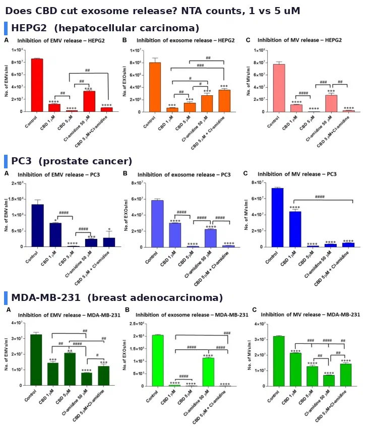
<p>The foundational paper is Kosgodage and colleagues, 2018, and its own title states the finding: CBD is a novel inhibitor for exosome and microvesicle release in cancer. Three cell lines, prostate PC3, hepatocellular HEPG2, breast adenocarcinoma MDA-MB-231. Exosome release dropped significantly in all three, dose-dependently at 1 and 5 micromolar, measured by nanoparticle tracking. Microvesicle release also fell, with more variability between lines. The paper ties the effect to mitochondrial function changes, modulation of STAT3 and prohibitin expression, and shows CBD sensitizing the cells to chemotherapy alongside the release inhibition <span class="evidence-badge evidence-badge-level" style="--evidence-color: #10b981" title="Level: AN. Confidence: HIGH.">AN · HIGH</span> for the in vitro finding. Three cell lines is a real replication across lineages, and it is still three immortal lines.</p>
<figure>
    <img loading="lazy" decoding="async"
         src="/cannabinoids-exosome-release/kosgodage-2018-exosome-release-three-lines_hu_5875e3a53c4d0d4d.webp"
         srcset="/cannabinoids-exosome-release/kosgodage-2018-exosome-release-three-lines_hu_4a7acd741b575f21.webp 640w"
         sizes="(max-width: 720px) 100vw, 720px"
         width="724" height="859"
         alt="Nanoparticle tracking analysis of vesicle release from three cancer cell lines treated with CBD at 1 and 5 micromolar for one hour: HEPG2 hepatocellular carcinoma, PC3 prostate cancer, and MDA-MB-231 breast adenocarcinoma, each showing reduced exosome release"></figure><p><em>The release finding as published, all three cell lines. Top: HEPG2. Middle: PC3. Bottom: MDA-MB-231. Particle counts by nanoparticle tracking, CBD at 1 and 5 micromolar, one hour. Reproduced from Kosgodage et al. 2018, Front Pharmacol 9:889, PMC6099119, CC-BY.</em></p>
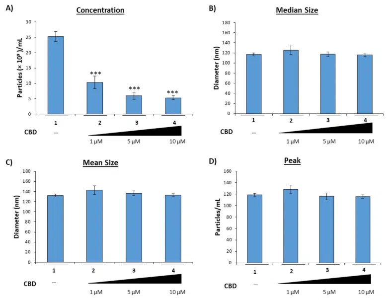
<p>DeMarino and colleagues, 2022, move the finding to infected cells, and this is the paper that matters most for this site's frame. In HIV-1 infected U1 monocytes and primary macrophages, CBD reduced the number of extracellular vesicles released, and the mechanism tracks two dials at once: reduced viral transcription and autophagy activation <span class="evidence-badge evidence-badge-level" style="--evidence-color: #f59e0b" title="Level: AN. Confidence: MODERATE.">AN · MODERATE</span>. The same group's context is why vesicles matter in infection: HIV RNAs such as TAR travel inside EVs and inflame recipient cells. Fewer vesicles, less cargo delivered. The observational hook in the same paper, cannabis use in people living with HIV associated with lower viral load, lower CD16+ monocytes and higher CD4 counts, is exactly that, observational, and it stays in its lane here.</p>
<figure>
    <img loading="lazy" decoding="async"
         src="/cannabinoids-exosome-release/demarino-2022-cbd-lowers-evs-u1_hu_ed046fe9f3aaefa.webp"
         srcset="/cannabinoids-exosome-release/demarino-2022-cbd-lowers-evs-u1_hu_a051d2e1c12a5cde.webp 640w"
         sizes="(max-width: 720px) 100vw, 720px"
         width="785" height="604"
         alt="CBD titration at 1, 5 and 10 micromolar in HIV-1 infected U1 monocytes over five days, showing reduced extracellular vesicle release by nanoparticle tracking analysis"></figure><p><em>The infected-cell version of the same experiment. HIV-1 infected U1 monocytes, CBD titrated at 1, 5 and 10 micromolar daily for five days, vesicle counts down. Reproduced from DeMarino et al. 2022, Cells 11(4):723, PMC8869966, CC-BY.</em></p>
<p>Under both sits the autophagy arm, and this is where the mechanism gets mapped residue by residue. Vrechi and colleagues, 2021, showed in human neuroblastoma SH-SY5Y and murine astrocyte lines that CBD-induced autophagy runs through CB1, CB2 and TRPV1, since receptor antagonists block it, through ERK1/2 activation and AKT suppression, and depends on the autophagy initiator ULK1 while being independent of mTORC1. That last combination is a non-canonical pathway, which matters for anyone arriving from the site's <a href="/spikeopathy-mtor-glymphatic-sirt1/">mTOR clearance piece</a>: CBD does not route through the mTOR lever that article documents, it runs a parallel one <span class="evidence-badge evidence-badge-level" style="--evidence-color: #10b981" title="Level: AN. Confidence: HIGH.">AN · HIGH</span>.</p>
<p>Wang and colleagues, 2022, take the arm to a whole organism, with the honest caveat that the organism is a worm. CBD extended C. elegans lifespan, promoted autophagic flux in nerve-ring neurons, and the extension vanished under RNAi knockdown of the autophagy genes bec-1, vps-34 and sqst-1, and under sir-2.1, the worm SIRT1 homolog. The same paper shows autophagic flux induction in mammalian hippocampal and SH-SY5Y neurons <span class="evidence-badge evidence-badge-level" style="--evidence-color: #f59e0b" title="Level: AN. Confidence: MODERATE.">AN · MODERATE</span>. And the group behind Vrechi 2021 published the follow-up in 2025: in tau-overexpressing SH-SY5Y cells, CBD at 100 nanomolar and 10 micromolar lowered total tau and phosphorylated AT8 tau through autophagy, with the autophagy blocker chloroquine reversing the effect <span class="evidence-badge evidence-badge-level" style="--evidence-color: #f59e0b" title="Level: AN. Confidence: MODERATE.">AN · MODERATE</span>. An autophagy inducer with a mapped mechanism, in the same mechanistic family as the site's SIRT1 material, is worth a reader's attention on its own terms, even before the vesicle story.</p>
<h2 id="the-entry-arm-adjacent-and-distinct">The entry arm, adjacent and distinct<a class="heading-anchor" href="#the-entry-arm-adjacent-and-distinct" aria-label="Link to this section">#</a></h2>
<p>Two cannabinoid papers usually get folded into this conversation that are doing something different, and the difference matters. Van Breemen and colleagues, 2022, used affinity selection mass spectrometry to find hemp cannabinoids that bind spike, and landed on the acids: cannabigerolic acid, CBGA, and cannabidiolic acid, CBDA. Both blocked infection of epithelial cells by pseudovirus and by live SARS-CoV-2, equally against alpha and beta variants, with micromolar affinities <span class="evidence-badge evidence-badge-level" style="--evidence-color: #10b981" title="Level: AN. Confidence: HIGH.">AN · HIGH</span> for the in vitro result. This is an entry-blockade arm, not a release arm. It says something about the virus getting in. It says nothing about vesicles getting out.</p>
<p>The second paper is the honesty test for the whole section. Anil and colleagues, 2021, tested a cannabis extract fraction high in CBD against COVID-relevant inflammation, and got a split result by cell type. In alveolar epithelial A549 cells, the fraction dose-dependently reduced IL-6, IL-8, the chemokines CCL2 and CCL7, and ACE2 expression. In macrophages, the same fraction increased IL-6 and IL-8 and phagocytosis, and a purified phytocannabinoid formulation flipped the macrophage result the other way <span class="evidence-badge evidence-badge-level" style="--evidence-color: #f59e0b" title="Level: AN. Confidence: MODERATE.">AN · MODERATE</span>. Same extract, opposite direction, different cell. The authors themselves attach the caution about proposing cannabis as a COVID treatment, and the finding deserves to travel with the citation. Any claim that cannabinoids are simply anti-inflammatory in this disease context dies on this paper.</p>
<h2 id="the-spike-side-of-the-ledger-and-the-untested-junction">The spike side of the ledger, and the untested junction<a class="heading-anchor" href="#the-spike-side-of-the-ledger-and-the-untested-junction" aria-label="Link to this section">#</a></h2>
<p>For the vesicle story to connect to this site's cluster, spike has to be on the shipping side of the transaction. Two papers put it there. Mishra and Banerjea, 2021, showed that cells transfected with spike release exosomes with reprogrammed cargo, loaded with miR-148a and miR-590, which recipient human microglia internalize; the microRNAs suppress USP33 and downstream IRF9 and flip the microglial cytokine profile toward TNF-alpha and NF-kB activation <span class="evidence-badge evidence-badge-level" style="--evidence-color: #f59e0b" title="Level: AN. Confidence: MODERATE.">AN · MODERATE</span>, a transfection model, stated as such. Frank and colleagues, 2022, showed the S1 subunit alone acts as a PAMP-like signal: injected intracisternally in rats it drove sickness behavior, and in vitro it activated microglia and TLR2 and TLR4 signaling directly <span class="evidence-badge evidence-badge-level" style="--evidence-color: #10b981" title="Level: AN. Confidence: HIGH.">AN · HIGH</span> for the receptor work.</p>
<p>So both halves of the transaction exist in the literature, each measured in its own system. Spike-bearing exosomes circulate in vaccinated humans by day 14 (Bansal, in the relay article's register). Stress and infected cells ship pro-inflammatory cargo in vesicles, and CBD turns vesicle release down in culture. The junction is the missing measurement, and it deserves its own sentence: no published study has tested whether cannabinoids modulate the release, cargo, or count of vaccine-derived or LNP-derived extracellular vesicles, in a cell model, in an animal, or in a person. The lever exists in one literature and the transport layer exists in another, and the experiment that puts them in the same room has not been run.</p>
<p>Here is the whole picture as one diagram, with the untested junction drawn as what it is, a dashed line:</p>
<div class="mermaid">

%%{init: {"flowchart": {"nodeSpacing": 30, "rankSpacing": 40}}}%%
flowchart TB
    A["Spike in producer cells"] --> B["Exosomal cargo reprogrammed"]
    B --> C["miR-148a and miR-590 loaded"]
    C --> D["Recipient microglia:<br/>USP33 and IRF9 down,<br/>TNF-alpha up"]
    A --> E["Spike-bearing exosomes<br/>in plasma by day 14"]
    F["CBD at 1 to 5 micromolar"] --> G["Exosome release down<br/>in three cancer lines"]
    F --> H["EV release down from<br/>HIV-1 infected myeloid cells"]
    F --> I["Autophagy up via ERK1/2,<br/>ULK1, SIRT1-dependent"]
    I --> J["Tau cleared in<br/>tau-overexpressing neurons"]
    K["CBGA and CBDA"] --> L["Spike binding blocked,<br/>entry blocked in vitro"]
    E -. "cannabinoid effect untested here" .-> F
    classDef ship fill:#e3ecf7,stroke:#5b7ea6,color:#1a1a2e
    classDef lever fill:#e7f0e3,stroke:#6a8f5a,color:#1a1a2e
    classDef entry fill:#f3ece0,stroke:#a08a5c,color:#1a1a2e
    class A,B,C,D,E ship
    class F,G,H,I,J lever
    class K,L entry

</div>

<p>Blue: the shipping side, what stressed and transfected cells release. Green: the release and autophagy levers. Tan: the separate entry arm. A dashed line is not a pathway. It is the shape of a gap.</p>
<h2 id="what-would-settle-it">What would settle it<a class="heading-anchor" href="#what-would-settle-it" aria-label="Link to this section">#</a></h2>
<p>The first experiment is unglamorous: take the DeMarino design and swap the infection for LNP transfection. Cells exposed to LNP-mRNA, vesicle counts by nanoparticle tracking, with and without CBD, and then the same question asked of the vesicle cargo. The second is the observational bridge: if any cohort of CBD users has serial vesicle measurements around vaccination, the hypothesis gets a real test without anyone dosing anybody. The third is the one that would end the argument either way, vesicle fractions from vaccinated plasma with and without cannabinoid exposure, assayed for spike and for vaccine ionizable lipid.</p>
<p>What would refute the interest: CBD failing to reduce vesicle release in primary human cells at concentrations achievable orally, or release inhibition that fails to change cargo transfer to recipient cells. Either result would collapse the junction without needing the human trial first.</p>
<h2 id="what-a-real-product-is-from-the-genomics-side">What a real product is, from the genomics side<a class="heading-anchor" href="#what-a-real-product-is-from-the-genomics-side" aria-label="Link to this section">#</a></h2>
<p>Everything above uses micromolar purified compound on cells. What people actually buy is where McKernan's cannabis material earns its place in this article, because the gap between the two is where the harm lives.</p>
<p>Most CBD on the market comes from Type III cannabis plants carrying an active CBDAS gene, which sets the flower's chemistry at roughly a 20:1 ratio of CBDA to THCA. As McKernan puts it in the Huberman follow-up post, most gummies are something like 20 milligrams of CBD with 1 milligram of THC riding along, and that accompanying THC is not nothing when the products are evaluated. Full-spectrum oils vary further, and parents in the epilepsy literature have reported longer seizure-reduction duration with them, which is consistent with entourage effects and impossible to bottle as a standard dose.</p>
<p>The interaction ceiling is the grapefruit rule: any drug carrying a grapefruit warning should be assumed to clear more slowly alongside high-dose CBD, because CBD inhibits the same CYP enzymes, CYP3A4 and CYP2C19 among them. McKernan's post names the concrete cases: clobazam, where the slowed-metabolism signal showed up in epilepsy trials, and warfarin and apixaban, both common in exactly the older populations using CBD for sleep and pain. The site's <a href="/spikeopathy-mtor-glymphatic-sirt1/">spikeopathy clearance piece</a> already carries the pharmacokinetics, around 6 percent oral bioavailability, multiplied by high-fat meals or piperine, and the CYP3A4 polypharmacy warning. This article adds the practitioner heuristic, not a dose.</p>
<p>And then the contamination findings, each from his own lab's measurements. Commercial flowers remediated by irradiation or ozone after a dirty grow can pass microbial tests while leaving endotoxin behind, undetected by the tests that exist. The largest cannabis microbiome program to date, Zamir Punja's, traced Aspergillus, Fusarium and Penicillium contamination in cocofibre growing medium through to flowering plants, seeds included, which undercuts the assumption that flower contamination is only an air-spread problem. His FOIA work with Yasha Kahn documented lab shopping, growers firing honest potency labs and hiring accommodating ones, replicated independently from Massachusetts regulator data. The hemp loophole rounds it out: 0.3 percent delta-9 THC compliance permits 24 percent THCA flower that converts to THC when burned, sold online with limited age checks. The site's supplement-writing rule, certificate of analysis or it does not exist, applies to this category with more force than almost any other.</p>
<p>One case deserves its own paragraph because of what it says about surveillance rather than chemistry. In 2025 a medical cannabis patient in Pennsylvania died of Cryptococcus neoformans, and the fungal genome from the dispensary product matched the patient's isolate at a handful of SNPs against tens of thousands separating it from wild strains. McKernan's post draws the surveillance conclusion this site has made about vaccine harm reporting, from the other direction: &quot;There is no VAERs for cannabis.&quot; Federal registries do not track cannabis fatalities, patients historically hid use to avoid CPS and organ-list consequences, so the familiar claim that no one has ever died from cannabis rests partly on no one keeping the ledger. The same under-tracking critique this site applies to one harm system applies to the other. Consistency is the whole job.</p>
<h2 id="what-this-article-is-not-claiming">What this article is not claiming<a class="heading-anchor" href="#what-this-article-is-not-claiming" aria-label="Link to this section">#</a></h2>
<ul>
<li>No claim that CBD clears spike, reduces vaccine-derived vesicles, or changes post-vaccine outcomes. The junction is untested and the takeaways say so.</li>
<li>No dosing claim. The culture experiments use micromolar purified compound; oral bioavailability is around 6 percent and product content is unreliable, which is a bigger barrier than any mechanism.</li>
<li>Infection evidence and vaccination evidence are kept separate throughout. DeMarino is HIV. Bansal is vaccination. Anil is neither, an inflammatory mimic, and it carries a direction reversal between cell types.</li>
<li>The worm and cell-line work is not human evidence. Kosgodage's three lines are immortal cancer lines, Wang's organism is a nematode, and the register marks every line accordingly.</li>
<li>The practitioner section is about product reality and interactions, not a recommendation. Anyone on warfarin, apixaban, clobazam or any grapefruit-flagged drug has a prescriber question to ask first.</li>
</ul>
<h2 id="what-would-confirm-it">What would confirm it<a class="heading-anchor" href="#what-would-confirm-it" aria-label="Link to this section">#</a></h2>
<ul>
<li>A replication of release inhibition in primary human cells, including monocyte-derived macrophages, at concentrations measured in plasma after oral dosing.</li>
<li>The LNP swap experiment: vesicle counts and cargo from LNP-exposed cells with and without cannabinoid treatment.</li>
<li>Any cohort evidence tying cannabinoid use to measurable differences in circulating vesicle counts or vesicle cargo after vaccination.</li>
</ul>
<h2 id="what-would-refute-it">What would refute it<a class="heading-anchor" href="#what-would-refute-it" aria-label="Link to this section">#</a></h2>
<ul>
<li>CBD failing to inhibit vesicle release in primary cells at achievable concentrations, which would confine the finding to immortal lines.</li>
<li>Release inhibition that leaves cargo transfer to recipient cells unchanged, which would make the mechanism irrelevant to the relay layer even if the count drops.</li>
<li>A well-powered observational cohort showing no difference in post-vaccination vesicle profiles between cannabinoid users and non-users.</li>
</ul>
<h2 id="citation-register-calibrated">Citation register (calibrated)<a class="heading-anchor" href="#citation-register-calibrated" aria-label="Link to this section">#</a></h2>
<p><strong>Release and autophagy arm</strong></p>
<ul>
<li>Kosgodage US, Mould R, Henley AB, et al. Cannabidiol (CBD) Is a Novel Inhibitor for Exosome and Microvesicle (EMV) Release in Cancer. <em>Front Pharmacol</em>. 2018;9:889. PMID 30150937. DOI 10.3389/fphar.2018.00889. PMC6099119. [AN, three cancer cell lines]</li>
<li>DeMarino C, Cowen M, Khatkar P, et al. Cannabinoids Reduce Extracellular Vesicle Release from HIV-1 Infected Myeloid Cells and Inhibit Viral Transcription. <em>Cells</em>. 2022;11(4):723. PMID 35203372. DOI 10.3390/cells11040723. PMC8869966. [AN, infected myeloid cells; observational associations noted in text]</li>
<li>Vrechi TAM, Leão AHFF, Morais IBM, et al. Cannabidiol induces autophagy via ERK1/2 activation in neural cells. <em>Sci Rep</em>. 2021;11:5434. PMID 33686185. DOI 10.1038/s41598-021-84879-2. PMC7940388. [AN, SH-SY5Y and astrocyte lines]</li>
<li>Wang Z, Zheng P, Chen X, et al. Cannabidiol induces autophagy and improves neuronal health associated with SIRT1 mediated longevity. <em>Geroscience</em>. 2022;44(3):1505-1524. PMID 35445360. DOI 10.1007/s11357-022-00559-7. PMC9213613. [AN, C. elegans plus mammalian neurons]</li>
<li>Vrechi TAM, Guarache GC, Oliveira RB, et al. Cannabidiol-Induced Autophagy Ameliorates Tau Protein Clearance. <em>Neurotox Res</em>. 2025;43(1):8. PMID 39900844. DOI 10.1007/s12640-025-00729-3. PMC11790692. [AN, tau-overexpressing SH-SY5Y]</li>
<li>Pereira GJDS, Leão AHFF, Erustes AG, et al. Pharmacological Modulators of Autophagy as a Potential Strategy for the Treatment of COVID-19. <em>Int J Mol Sci</em>. 2021;22(8):4067. PMID 33920748. DOI 10.3390/ijms22084067. PMC8071111. [review, positions CBD in the autophagy-modulator set]</li>
</ul>
<p><strong>Entry arm</strong></p>
<ul>
<li>van Breemen RB, Muchiri RN, Bates TA, et al. Cannabinoids Block Cellular Entry of SARS-CoV-2 and the Emerging Variants. <em>J Nat Prod</em>. 2022;85(1):176-184. PMID 35007072. DOI 10.1021/acs.jnatprod.1c00946. PMC8768006. [AN, pseudovirus plus live virus]</li>
<li>Anil SM, Shalev N, Vinayaka AC, et al. Cannabis compounds exhibit anti-inflammatory activity in vitro in COVID-19-related inflammation in lung epithelial cells and pro-inflammatory activity in macrophages. <em>Sci Rep</em>. 2021;11:1462. PMID 33446817. DOI 10.1038/s41598-021-81049-2. PMC7809280. [AN, direction reversal by cell type]</li>
</ul>
<p><strong>Spike shipping side</strong></p>
<ul>
<li>Mishra R, Banerjea AC. SARS-CoV-2 Spike Targets USP33-IRF9 Axis via Exosomal miR-148a to Activate Human Microglia. <em>Front Immunol</em>. 2021;12:656700. PMID 33936086. DOI 10.3389/fimmu.2021.656700. PMC8079643. [AN, transfection model]</li>
<li>Frank MG, Nguyen KH, Ball JB, et al. SARS-CoV-2 spike S1 subunit induces neuroinflammatory, microglial and behavioral sickness responses: Evidence of PAMP-like properties. <em>Brain Behav Immun</em>. 2022;100:267-277. PMID 34915155. DOI 10.1016/j.bbi.2021.12.007. PMC8667429. [AN, rat ICM plus receptor work]</li>
<li>Bansal S, et al. Cutting Edge: Circulating exosomes with COVID spike protein are induced by BNT162b2 mRNA vaccination. <em>J Immunol</em>. 2021;207(10):2405-2410. PMID 34654691. DOI 10.4049/jimmunol.2100637. [PP, small cohort; full treatment in the relay article]</li>
</ul>
<p><strong>McKernan's posts, practitioner material, secondary not primary</strong></p>
<ul>
<li>Huberman speaks with Matt Hill on Cannabinoids, Nepetalactone Newsletter, 2024: <a href="https://anandamide.substack.com/p/huberman-speaks-with-mark-hill-on">link</a>. Source for the CBDA:THCA ratio, the grapefruit rule, clobazam and anticoagulant cases, biphasic dosing.</li>
<li>High levels of Endotoxin on commercial cannabis flowers, 2023: <a href="https://anandamide.substack.com/p/high-levels-of-endotoxin-on-commercial">link</a>. Source for remediation leaving endotoxin.</li>
<li>Big Cannabis behaving like Big Tobacco, 2024: <a href="https://anandamide.substack.com/p/big-cannabis-behaving-like-big-tobacco">link</a>. Source for lab shopping, the FOIA replication, his conflict statement, the 2011 genome.</li>
<li>Cocofibre can be the source of fungal contamination in Cannabis, 2025: <a href="https://anandamide.substack.com/p/cocofibre-can-be-the-source-of-fungal">link</a>. Source for the Punja microbiome program and the cocofibre transmission route.</li>
<li>Cryptococcus neoformans brings a fatality to the medical Cannabis industry, 2025: <a href="https://anandamide.substack.com/p/cryptococcus-neoformans-brings-a">link</a>. Source for the Pennsylvania case and the surveillance asymmetry.</li>
</ul>
<h2 id="open-to-corrections">Open to corrections<a class="heading-anchor" href="#open-to-corrections" aria-label="Link to this section">#</a></h2>
<p>Every number here traces to the register above, and the one claim people will want to make, that CBD cleans up the vesicle layer, is exactly the claim the evidence does not support yet. The culture findings are real and replicated in direction across systems. The junction is unmeasured. If something here is wrong, tell me and it gets corrected with the same prominence as the original claim. General policy on the <a href="/methodology/">Methodology page</a>.</p>
]]></content></entry><entry><title>The Exosome Relay: Spike Without a Factory</title><link rel="alternate" href="https://measslainte.com/the-exosome-relay/"/><id>https://measslainte.com/the-exosome-relay/</id><published>2026-08-30T00:00:00+01:00</published><updated>2026-10-06T19:57:06+01:00</updated><summary type="html">A hypothesis-graded mechanism piece: how a short-lived transfected cell, durable membrane-bound protein, and extracellular-vesicle transport can explain long-term spike detection without genomic integration. Frame credited to Maria Gutschi; every load-bearing claim cited to primary literature; the patisiran lipid half-life corrected to the published 14.6-28.7 days.</summary><content type="html"><![CDATA[<h2 id="why-this-article-exists">Why this article exists<a class="heading-anchor" href="#why-this-article-exists" aria-label="Link to this section">#</a></h2>
<p>There is a standing objection to everything this site documents about persistent spike. It runs: modified mRNA degrades within days, free protein clears within weeks, so spike detected months after injection implies either genomic integration or a chronic producer, meaning infection or something like it. Both implications are serious. Neither is required.</p>
<p>A third explanation exists, each of whose layers is already in the published literature: the transfected cell is finite, the protein it made is durable, and the cell's own vesicle machinery can carry lipid, RNA fragments and protein out to other sites. The frame for putting those three layers together is not mine. It belongs to a pharmacist, Maria Gutschi, writing as @CanningPharm. This article holds her frame against the primary literature, corrects two numbers that usually travel with it, and wires it into this site's persistence cluster.</p>
<p>Tagged HYPOTHESIS. The layers underneath are graded separately, and the grading matters: what is measured, what is analogous, and what is proposed are kept apart throughout.</p>
<h2 id="credit-where-it-belongs">Credit where it belongs<a class="heading-anchor" href="#credit-where-it-belongs" aria-label="Link to this section">#</a></h2>
<p>The Index / Persistence / Relay frame is Gutschi's, published in her thread (<a href="https://x.com/CanningPharm/status/2093523693687193785">X, @CanningPharm</a>) and expanded in her Substack essay (<a href="https://mariagutschi.substack.com/p/the-exosome-problem-why-the-mrna">The Exosome Problem</a>). Nothing in this article modifies or extends her model. What this article adds is the site's standard treatment: primary sourcing for every load-bearing claim, calibration of the two figures that get quoted loosely, explicit refutation criteria, and crosslinks into the persistence articles this site already carries.</p>
<h2 id="layer-1-the-index-cell-a-finite-template">Layer 1: the index cell, a finite template<a class="heading-anchor" href="#layer-1-the-index-cell-a-finite-template" aria-label="Link to this section">#</a></h2>
<p>The first transfected cell receives a finite amount of modified mRNA. Early public statements put its life at a few days. The measured picture is longer, and still bounded.</p>
<p>In axillary lymph-node biopsies after mRNA vaccination, Röltgen and colleagues detected vaccine mRNA, and spike protein, in germinal centres out to 60 days <span class="evidence-badge evidence-badge-level" style="--evidence-color: #10b981" title="Level: PP. Confidence: HIGH.">PP · HIGH</span> for the detection finding itself, a small biopsy series. Sixty days is roughly an order of magnitude beyond the few-days figure, and it is not forever.</p>
<p>The plasma kinetics agree. Ogata and colleagues, using ultrasensitive Single Molecule Array assays in 13 recipients of mRNA-1273, found circulating S1 in 11 of 13 participants from day 1 after the first dose, peaking around day 5, no longer detectable by day 14, and absent after the second dose <span class="evidence-badge evidence-badge-level" style="--evidence-color: #f59e0b" title="Level: PP. Confidence: MODERATE.">PP · MODERATE</span>, n=13. Antigen appearance in plasma is early, and its clearance tracks the rise of the antibody response.</p>
<p>Layer 1 is a real but bounded template. <span class="evidence-badge evidence-badge-level" style="--evidence-color: #f59e0b" title="Level: MECHANISTIC. Confidence: MODERATE.">MECHANISTIC · MODERATE</span> as a general statement, carried by the two human series above.</p>
<h2 id="layer-2-persistence-protein-after-the-template">Layer 2: persistence, protein after the template<a class="heading-anchor" href="#layer-2-persistence-protein-after-the-template" aria-label="Link to this section">#</a></h2>
<p>The same two papers carry the second layer. In Ogata's series, plasma S1 is present while the template is being consumed: protein, once made, exists on a timescale of its own. In Röltgen's biopsies, spike protein sits in germinal centres alongside detectable mRNA at 60 days, that is, deposited antigen in tissue, not circulating antigen in plasma.</p>
<p>The site's persistence cluster documents the downstream of this: durable antigen display, reservoirs, and the failure of clearance that lets it matter clinically. This layer is where the relay article meets that cluster, and the crosslinks at the end of this article are that connection made explicit.</p>
<h2 id="layer-3-the-relay-out-through-the-vesicle-machinery">Layer 3: the relay, out through the vesicle machinery<a class="heading-anchor" href="#layer-3-the-relay-out-through-the-vesicle-machinery" aria-label="Link to this section">#</a></h2>
<p>This is the layer the objection never accounts for, and it is the best-measured of the three.</p>
<p>The quantitative baseline comes from siRNA-LNP work: of internally delivered LNP cargo, roughly 1-2% reaches the cytosol, while around 70% is exocytosed back out of the cell through recycling endosomes and multivesicular bodies <span class="evidence-badge evidence-badge-level" style="--evidence-color: #10b981" title="Level: AN. Confidence: HIGH.">AN · HIGH</span> for the in vitro quantification. The dominant fate of endocytosed LNP contents is exit, and the exit route is the same machinery that manufactures exosomes.</p>
<p>Maugeri and colleagues then showed the exit is functional, for an LNP-mRNA product in vitro: mRNA and ionizable lipid that had escaped endosomes were loaded into extracellular vesicles and released, and vesicle-delivered mRNA was translated in recipient cells <span class="evidence-badge evidence-badge-level" style="--evidence-color: #10b981" title="Level: AN. Confidence: HIGH.">AN · HIGH</span>. Ionizable lipid travels in the vesicles. That single point does a lot of work, and it is measured, in that system.</p>
<p>And in vaccinated humans, Bansal and colleagues found circulating exosomes carrying spike protein by day 14 after the first BNT162b2 dose, with anti-spike antibodies appearing on the same timescale after the second <span class="evidence-badge evidence-badge-level" style="--evidence-color: #f59e0b" title="Level: PP. Confidence: MODERATE.">PP · MODERATE</span>. The relay is not only a culture phenomenon. Vesicle-borne spike is observable in people.</p>
<p>The three layers as one diagram, redrawn from the frame above rather than linked from it:</p>
<div class="mermaid">

flowchart LR
    A["Index cell: finite modified mRNA"] --> B["Translation: spike protein"]
    A --> C["Endosome / multivesicular body"]
    C -->|"1-2% escapes to cytosol"| D["Cytosolic mRNA, translated"]
    C -->|"~70% recycled outward"| E["Exosomes: lipid, RNA fragments, spike"]
    B --> F["Spike on membranes and in tissue, durable"]
    B --> G["Plasma S1: days 1-14, then cleared"]
    E --> H["Recipient cells at distant sites"]
    H --> I["Translated or displayed spike, antigen presented"]

</div>

<p>An LNP dose adds one thing natural infection does not: a bulk deposit of membrane-active ionizable lipid into the endosomal system of the transfected cell, the same compartment that ships vesicles. Maugeri's ionizable-lipid-in-EV finding is the reason this difference is more than hand-waving. Whether vaccine-derived EVs in humans carry ALC-0315 or SM-102 inside them has not been measured; that distinction between infection-generated and LNP-generated vesicles is proposed, and the confirmation section below names the measurement that would settle it.</p>
<h2 id="the-closest-licensed-analog-calibrated">The closest licensed analog, calibrated<a class="heading-anchor" href="#the-closest-licensed-analog-calibrated" aria-label="Link to this section">#</a></h2>
<p>Gutschi's essay leans on Onpattro (patisiran), the one licensed LNP RNA drug with full public pharmacokinetics, as the analog for what cells do with internalized LNP. The published clinical PK supports a biphasic-plus picture: rapid distribution clearance, then a minor secondary peak, then a long terminal phase, with the ionizable lipid DLin-MC3-DMA showing a terminal half-life of 14.6 to 28.7 days, against 3.2 ± 1.8 days for the siRNA itself <span class="evidence-badge evidence-badge-level" style="--evidence-color: #10b981" title="Level: PP. Confidence: HIGH.">PP · HIGH</span> for the PK as published. A secondary peak is what redistributed lipid coming back out of tissue looks like, which is at least consistent with cells taking LNP up and sending components back out.</p>
<p>Two calibrations, because the numbers that circulate with this frame are looser than their sources:</p>
<ol>
<li>The ~60-day lipid half-life quoted in the essay is not what the primary source says. The published terminal half-life of DLin-MC3-DMA in humans is 14.6 to 28.7 days. This article carries the published range.</li>
<li>The 70% re-export figure is Sahay's in vitro siRNA-LNP quantification, not a clinical Onpattro measurement. It is the right order for the argument, and it is in vitro.</li>
</ol>
<p>The analogy itself is graded [HYPOTHESIS], LOW-MODERATE: patisiran is an intravenous siRNA product cleared into hepatocytes, the vaccines are intramuscular mRNA products draining to node and tissue. Different cargo, different route, different organ. The analog tells you what cells can do with internalized LNP. It does not tell you what these products did.</p>
<h2 id="what-the-model-explains">What the model explains<a class="heading-anchor" href="#what-the-model-explains" aria-label="Link to this section">#</a></h2>
<ul>
<li>Detection without integration. The site's insertional-mutagenesis defence piece is careful about integration claims. This frame is the mechanism that does the work integration is usually invoked for: persistence of antigen without any change to the genome. <a href="/insertional-mutagenesis-defense/">insertional-mutagenesis defence</a></li>
<li>Distribution. Antigen and lipid found far from the injection site and its draining nodes do not require cells to migrate. Vesicles go with the circulation.</li>
<li>Re-seeding during the tolerance window. The tolerance-gate article asks what would confirm a window in which antigen is detected loudly and cleared quietly. Antigen arriving in vesicles while the gate is open is exactly that observation class. <a href="/why-spike-persists-tolerance-gate/">why spike persists: the tolerance gate</a></li>
<li>Persistent antigenic drive. The persistence cluster documents that spike stays detectable and that clearance fails. The relay supplies a way for presentation to continue after the producer cell is gone. <a href="/spike-persistence-microclots-reactivated-viruses/">spike persistence, microclots, reactivated viruses</a></li>
</ul>
<h2 id="what-this-article-is-not-claiming">What this article is not claiming<a class="heading-anchor" href="#what-this-article-is-not-claiming" aria-label="Link to this section">#</a></h2>
<ul>
<li>No integration claim. The frame works precisely because integration is not needed.</li>
<li>No immortal factory. The index cell is finite; on this model the vesicle output is bounded by what the index cell and its neighbours took up, minus clearance.</li>
<li>No dosimetry. Nothing here converts vesicle counts into any quantity per dose.</li>
<li>The infection-EV versus LNP-EV cargo difference is proposed, not established. One half of it, ionizable lipid travelling in EVs, is measured in vitro (Maugeri). The other half, vaccine-derived EVs carrying ALC-0315 or SM-102 in humans, is measured nowhere yet.</li>
<li>Small-n primaries are stated as such. Ogata is 13 participants, Bansal a small longitudinal cohort, Röltgen a biopsy series. Peer-reviewed human data with small n is still small n, and the register below says so at every line.</li>
</ul>
<h2 id="what-would-confirm-it">What would confirm it<a class="heading-anchor" href="#what-would-confirm-it" aria-label="Link to this section">#</a></h2>
<ul>
<li>Isolation of EV fractions from vaccinated plasma containing the vaccine ionizable lipids, ALC-0315 or SM-102, by mass spectrometry.</li>
<li>EV RNA sequencing showing vaccine-sequence RNA in vesicle fractions weeks after dosing.</li>
<li>A time course in which vesicle-borne antigen persists on the protein's timescale rather than the template's.</li>
</ul>
<h2 id="what-would-refute-it">What would refute it<a class="heading-anchor" href="#what-would-refute-it" aria-label="Link to this section">#</a></h2>
<ul>
<li>Competent EV isolation across cohorts finding neither vaccine-sequence RNA nor spike in vesicle fractions.</li>
<li>Kinetics in which vesicle-borne antigen tracks the template and disappears with it, which would make the relay a shadow of layer 1 rather than a mechanism of its own.</li>
</ul>
<h2 id="citation-register-calibrated">Citation register (calibrated)<a class="heading-anchor" href="#citation-register-calibrated" aria-label="Link to this section">#</a></h2>
<ul>
<li>Ogata AF, Cheng C-A, Desjardins M, et al. Circulating SARS-CoV-2 vaccine antigen detected in the plasma of mRNA-1273 vaccine recipients. <em>Clin Infect Dis</em>. 2022;74(4):715-718. PMID 34015087. DOI 10.1093/cid/ciab465. PMC8241425. [PP, n=13]</li>
<li>Röltgen K, et al. Immune imprinting, breadth of variant recognition, and germinal center response in human SARS-CoV-2 infection and vaccination. <em>Cell</em>. 2022;185(6):1025-1040. PMID 35148837. DOI 10.1016/j.cell.2022.01.018. [PP, biopsy series]</li>
<li>Maugeri M, et al. Linkage between endosomal escape of LNP-mRNA and loading into EVs for transport to other cells. <em>Nat Commun</em>. 2019;10:4333. PMID 31551417. DOI 10.1038/s41467-019-12275-6. [AN, in vitro]</li>
<li>Sahay G, et al. Efficiency of siRNA delivery by lipid nanoparticles is limited by endocytic recycling. <em>Nat Biotechnol</em>. 2013;31:653-658. PMID 23792629. DOI 10.1038/nbt.2614. PMC3814166. [AN, in vitro quantification]</li>
<li>Bansal S, et al. Cutting Edge: Circulating exosomes with COVID spike protein are induced by BNT162b2 mRNA vaccination. <em>J Immunol</em>. 2021;207(10):2405-2410. PMID 34654691. DOI 10.4049/jimmunol.2100637. [PP, small cohort]</li>
<li>Zhang X, et al. Pharmacokinetics of patisiran, the first approved RNA interference therapy, in patients with hereditary transthyretin-mediated amyloidosis. <em>J Clin Pharmacol</em>. 2020. PMID 31777097. DOI 10.1002/jcph.1553. PMC7187331. [PP, clinical PK]</li>
<li>Gutschi M. The exosome frame: Index, Persistence, Relay. Thread, X/@CanningPharm, 2026: <a href="https://x.com/CanningPharm/status/2093523693687193785">link</a>. Essay: <a href="https://mariagutschi.substack.com/p/the-exosome-problem-why-the-mrna">link</a>. [secondary frame, not a primary source]</li>
</ul>
<h2 id="open-to-corrections">Open to corrections<a class="heading-anchor" href="#open-to-corrections" aria-label="Link to this section">#</a></h2>
<p>Every number here is traceable to the register above, and the two corrections to the circulating figures are stated in the analog section, not buried. If something here is wrong, tell me and it gets corrected with the same prominence as the original claim. General policy on the <a href="/methodology/">Methodology page</a>.</p>
<hr>
<p><em>End of exosome relay article. The HYPOTHESIS tag comes off the relay layer the day someone publishes ALC-0315 or SM-102 inside EV fractions from vaccinated plasma. The refutation criteria above are the other way out.</em></p>
]]></content></entry></feed>