Table of Contents
Key Takeaways
- The brain has two waste-removal systems: intracellular autophagy and interstitial glymphatic flushing; both are energy-dependent and run mainly in deep sleep.
- Spike protein may damage both by impairing mitochondria: waste accumulates inside and between cells, inflammation rises, sleep worsens, and the cycle feeds itself.
- The broadest-reach levers are deep sleep, time-restricted eating, daily movement, and mitochondrial and clearance-supporting compounds.
- This is one model among several: testable, evidence-fitting, and not proven.
Where this sits. This article is one node of a larger model: spikeopathy as systemic clearance and tolerance failure under persistent antigenic drive. The model, the full lever map, and how the pieces connect live on the Spikeopathy hub.
Plain English#
Your brain has two waste-removal systems. One cleans the inside of cells, like taking out the rubbish. One flushes the spaces between cells, like a kitchen drain. Both need energy. Both run mainly while you sleep.
Spike protein (from the virus or the vaccine) may damage both. It hurts mitochondria, the cell's power supply. Without power, the rubbish piles up inside cells and the drain clogs between them. Waste builds up, inflammation rises, sleep gets worse, and the cycle feeds itself. That's the brain fog and exhaustion many people report.
This is one model among several. It is not proven. But it is testable, it fits the evidence, and, importantly, it points to things you can actually do.
You are not powerless here. The levers with the broadest mechanistic reach are simple: deep sleep, time-restricted eating, daily movement, and a handful of compounds that support mitochondria and clearance. None of this is a cure. All of it is within reach. The full article explains the biology and the evidence, so you can decide, with your doctor, what's worth trying.
What you can actually do#
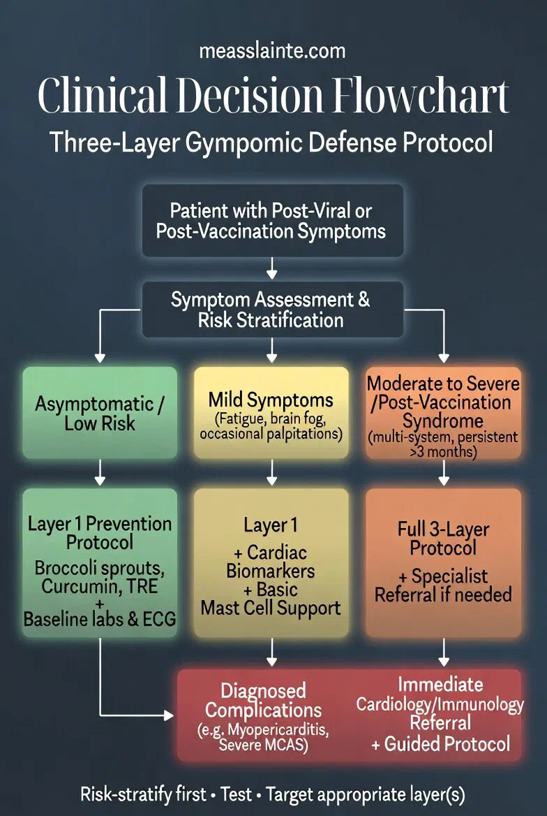
Grouped by which part of the cycle each lever touches. Not ranked. Different people will need different entry points. None of this is medical advice. Work with a clinician, especially if you are on prescription medication, pregnant, or managing a chronic condition.
1. Sleep: the glymphatic system runs mainly in deep sleep. This is the single most evidence-backed lever for brain waste clearance. Protect the last hour before bed (no screens, dim light), consistent sleep/wake times, cool dark room. If sleep is fragmented (common in post-COVID syndromes), address it first. Low-dose melatonin (0.3–1 mg, 1–2 hours before bed) is mechanistically reasonable for sleep-gated clearance [ESTABLISHED for sleep/glymphatic; MECHANISTIC for melatonin dose].
2. Time-restricted eating: the body's natural cleanup switch for inside cells. Eating within an 8–10 hour window (e.g. 9am–5pm or 10am–6pm) triggers autophagy, the process that clears damaged proteins and mitochondria. Start with 12 hours overnight and tighten gradually. Not suitable for everyone: underweight, pregnant, eating-disorder history, or uncontrolled diabetes need clinician input [ESTABLISHED for autophagy induction; MECHANISTIC for spikeopathy application].
3. Daily movement: drives the glymphatic pump. Arterial pulsation and breathing both pump CSF through the brain. Walking, cycling, anything that raises heart rate modestly. 30 minutes most days. If you have post-exertional malaise (PEM), common in long COVID, this must be paced, not pushed. Pushing through PEM makes things worse. If exercise crashes you, stop and consult a clinician familiar with ME/CFS or PASC [ESTABLISHED for glymphatic pumping; PEM caveat is clinical consensus].
4. Mitochondrial support: the energy supply both systems depend on. The compounds with the cleanest mechanistic case here are NAD⁺ precursors (NR or NMN), ubiquinol (reduced CoQ10), and PBM (near-infrared / red light therapy). These target the SIRT1/PGC-1α axis and electron transport, the energy backbone. See the Tier 1 matrix below for details and dosing cautions [MECHANISTIC; small HUMAN pilot signal for PBM in long-COVID cognitive symptoms].
5. Inflammation modulation: calms the cytokines that mislocalise AQP4. Sulforaphane (broccoli sprouts) activates Nrf2. Baicalin (Chinese skullcap) inhibits cGAS-STING and is antiviral against SARS-CoV-2 3CL protease in vitro. CBD has the strongest spike-specific in vitro evidence of the cannabinoids (Corpetti 2021, PPARγ → TLR4/NLRP3 suppression), but no human trials for these syndromes, and significant CYP3A4 drug-interaction risk. For the meningeal mast-cell arm (Theoharides 2026, see Pathway 2), mast-cell stabilisers (luteolin, quercetin, ketotifen, cromolyn, low-dose naltrexone) form a separate lever class worth flagging with your clinician if histamine/mast-cell symptoms (flushing, food sensitivities, POTS overlap) are part of your picture. Quality sourcing matters; see Tier 2 matrix [MECHANISTIC; no human RCTs for PASC/PVS].
6. Microclot / fibrin clearance: if the plumbing is clogged, clearance can't flow. Nattokinase and lumbrokinase are fibrinolytic enzymes with small human pilot data. This is the lever most likely to interact with anticoagulants and most needs clinician oversight: do not self-prescribe if on blood thinners. Full biomarker panel and cautions in the Spike Persistence companion [MECHANISTIC + HUMAN (small pilots)].
Baseline labs worth asking your clinician about (not a complete list, see Spike Persistence biomarker panel): D-dimer, fibrinogen, hs-CRP, ferritin, homocysteine, NAD⁺ (research-grade), CBC, metabolic panel. Track symptoms alongside any intervention; symptom logs beat memory every time.
What to avoid: pushing through crashes (PEM is real and worsens outcomes), polypharmacy without a clinician reviewing interactions (CBD, piperine, berberine, grapefruit all hit CYP3A4), and any source promising a "cure" for long COVID or post-vaccine syndrome. There is no cure yet. Anyone claiming otherwise is wrong or selling something. Move on.
Evidence protocol. Every claim gets a tag.
[ESTABLISHED]= multiple RCTs or strong human data.[MECHANISTIC]= in vitro or animal, pathway level.[HUMAN-IMAGING]= clinical cohorts with imaging or biomarkers.[AUTOPSY]= tissue series.[HYPOTHESIS]= convergent inference, not yet confirmed at scale. Weigh each claim on its own. Don't bundle them. Full grading system, sourcing standard, and correction policy on the Methodology page.
How this post fits in the series#
This is the pathway mechanics companion. It fills the gap between two posts I've already written. The triangle looks like this:
| Question | Read this | What it covers |
|---|---|---|
| Why won't spike clear? | Spike Persistence: Microclots, Reactivated Viruses | Tissue reservoirs, mTOR/p53 survival mechanism, RAGE/IL-10 tolerance trap, TMEM106B, microclots, APOE4/BBB, full biomarker panel |
| What compounds work? | Spike Protocol: Evidence Snapshot | Nattokinase, NAC, curcumin, quercetin, lions mane, oregano. Evidence tiers, dosing cautions, lab monitoring |
| What clearance systems fail? | You are here. | The glymphatic system (missing from both posts above). The SIRT1/PGC-1α axis that ties intracellular and extracellular clearance together. The dual-mTOR model. The device and compound levers unique to these pathways |
What's genuinely new here, not covered elsewhere on the site: the glymphatic dimension, SIRT1/PGC-1α as the connecting axis, the dual-mTOR model, PBM/NIR, HBOT, ubiquinol, urolithin A. For mTOR/p53 basics, spermidine, rapamycin, and the full coagulation/inflammation biomarker panel, follow the links above. I'm not re-explaining them here.
Executive Summary#
This post explores the clearance-system failure hypothesis of spikeopathy. It's one of several competing models for post-COVID and post-vaccination syndromes, not the only one. It's the pathway-mechanics companion to two existing posts (Spike Persistence and Spike Protocol) and it adds what neither covers: the glymphatic dimension.
The central model. Two energy- and inflammation-sensitive clearance systems fail together in chronic spikeopathy, and they reinforce each other:
- Intracellular clearance runs through the mTOR / SIRT1 / PGC-1α axis. mTORC1 hyperactivation suppresses autophagy. Misfolded proteins (amyloid-β, tau, α-synuclein) and damaged mitochondria accumulate.
- Extracellular clearance runs through the glymphatic system. Neuroinflammation disrupts astrocytic AQP4 polarisation and sleep-dependent waste removal. Proteins and metabolites back up.
A third upstream arm (covered in Pathway 2): the meningeal mast-cell arm (Theoharides 2026). Spike activates meningeal mast cells via TLR4/ACE2, releasing tryptase, histamine, IL-1β, IL-33, disrupting the BBB, and creating a mast-cell–microglia loop that sits upstream of both clearance systems above. Proposed, not proven in humans. Covered because it identifies a distinct lever class (mast-cell stabilisers) the other two arms don't.
Shared upstream driver. Spike-driven mitochondrial dysfunction and inflammation disrupt the SIRT1/PGC-1α axis. Both clearance systems lose their energy supply. The cycle amplifies.
Three supporting pillars:
- 2025 human imaging evidence
[HUMAN-IMAGING]: DTI-ALPS studies (He et al., Frontiers in Psychology) show glymphatic disruption tracking cognition and fatigue in COVID-recovered patients. The Cog-PASC multimodal study (Seo, Choi et al., Nature Communications) documents reduced clearance, white-matter injury, and elevated GFAP/NfL. - The dual-mTOR model
[HYPOTHESIS]: context-dependent mTOR hyperactivation (supporting viral persistence in reservoirs) alongside relative suppression elsewhere. This is why spikeopathy presentations are heterogeneous rather than uniform. - The cGAS-STING bridge
[MECHANISTIC]: plasmid DNA contamination and spike-driven mitochondrial damage both feed cytosolic DNA into cGAS-STING. That drives chronic type I IFN plus NF-κB, neuroinflammation, AQP4 mislocalisation, glymphatic failure. This connects the DNA-contamination angle (sequence analysis of the BNT162b2 plasmid, documented elsewhere on this site) directly to the clearance-failure angle.
What this post does NOT do. It doesn't claim any intervention reverses spikeopathy. It doesn't re-cover lab origin, DNA contamination forensics, oncogenic p53/RAGE pathways, or regulatory failure. Those live in genomic-defense-executive-summary, DNA-Contamination, hiv-protein-mimicry, and spike-persistence respectively. This is the clearance-mechanics layer only.
Scientific accountability. The model makes specific, falsifiable predictions (see Falsifiability section). If DTI-ALPS does not respond to clearance-enhancing interventions in future trials, or if SIRT1/PGC-1α signatures do not predict symptom trajectory, the hypothesis should be revised or abandoned. Not defended.
Background: Defining Spikeopathy#
A note on terminology. "Spikeopathy" is a working hypothesis term used in independent research communities. It's not a mainstream clinical diagnosis. It doesn't appear in ICD-10/11, NICE guidelines, or WHO classifications. Mainstream neurology and immunology currently frame post-COVID and post-vaccination symptoms as multifactorial syndromes (long COVID, PASC, PVS) with several competing models: viral persistence, autoimmunity, mitochondrial dysfunction, dysautonomia, microbiome disruption, others. This post explores one of those models, the clearance-system failure hypothesis, as a mechanistic framework. It is not a claim that this model is confirmed, complete, or superior to alternative explanations. Read it as a structured exploration of a hypothesis, not a clinical diagnosis guide.
Spikeopathy refers to chronic sequelae mechanistically linked to persistent SARS-CoV-2 spike protein, whether from infection or mRNA vaccination. Core features: neuroinflammation, protein misfolding and aggregation, mitochondrial dysfunction, endothelial damage, and symptoms like brain fog, fatigue, autonomic issues, with theoretical elevation in neurodegeneration risk.
The term is mechanistic, not a claim of confirmed population-level prion or dementia epidemic. As of mid-2026, evidence remains mechanistic plus imaging plus tissue-level, not definitive epidemiological causation [ESTABLISHED]. Surveillance gaps could miss delayed, heterogeneous signals.
Limits of the persistence hypothesis#
Honest constraints on the central premise:
- Not universal. Spike protein, S1 subunit, or mRNA components have been detected in some, not all, long COVID / PASC / PVS cases, in circulation, monocytes, and tissues (meninges, skull marrow, brain) up to months or years post-exposure. Persistence is documented in subsets. Some patients with detectable spike are asymptomatic.
- Causation unproven. Correlation exists in subsets, but whether persistence drives symptoms or is a bystander is not established. Confounders include reinfection, comorbidities, genetic susceptibility (e.g. ApoE4), and assay limits (distinguishing infection-derived vs vaccine-derived spike).
- Multifactorial syndromes. PASC and PVS have several competing models: viral persistence, autoimmunity, mitochondrial dysfunction, microclots, dysautonomia, microbiome disruption. Not every case fits a single mechanism. The clearance-system failure framework in this post is one of those models, not the umbrella.
- No direct human data for the EPO-EPOR-spike link. The Zhang et al. 2025 cDC1/EPOR findings are from mouse tolerance/allograft/tumour models. No published study directly tests EPO elevation or EPOR signalling in human spike persistence cohorts. Application to spikeopathy is mechanistic inference, not established.
- Persistence may involve multiple evasion strategies. Immune-privileged sites, microclot-impaired clearance, and broader immunosuppression are parallel candidates, not mutually exclusive with the EPOR/tolerance angle.
Frame the model as one piece of a complex puzzle, not a complete explanation.
Pathway 1: The SIRT1/PGC-1α Axis: What Connects the Two Clearance Systems#
Baseline mTOR/spike survival mechanism. The full mTORC1/autophagy biology, the Melo et al. 2025 mTOR/p53 model, rapamycin evidence, and the spermidine/resveratrol/EGCG compound details are in Spike Persistence: Microclots, Reactivated Viruses → Therapeutic Implications: Targeting mTOR & Autophagy. This post focuses on what's missing from that picture: the upstream axis that ties intracellular clearance to extracellular clearance.
Why SIRT1/PGC-1α is the critical node#
The standard mTOR story (spike → mTORC1 hyperactivation → autophagy suppressed → aggregates accumulate) is correct but incomplete. It doesn't explain why mTOR stays hyperactive, why mitochondria fail, or why the glymphatic system breaks down in parallel. The connecting axis is SIRT1 / PGC-1α:
- SIRT1 (NAD⁺-dependent deacetylase) is the cellular energy sensor. When NAD⁺ is abundant, SIRT1 is active.
- PGC-1α is SIRT1's primary target. It's the master regulator of mitochondrial biogenesis, fatty-acid oxidation, and mitophagy
[ESTABLISHED]. - Active SIRT1/PGC-1α restrains mTORC1 (via AMPK) and promotes autophagy
[ESTABLISHED]. - The same energy state also drives glymphatic pulsation: arterial pulsatility, AQP4 polarisation, CSF flow. All ATP-dependent.
The dual-mTOR model [HYPOTHESIS]: spike protein disrupts SIRT1/PGC-1α directly, through NAD⁺ depletion, inflammation-driven ACSS2/poly-ADP-ribosylation, and mitochondrial damage. This single hit cascades into:
- mTORC1 derepression, autophagy failure, intracellular waste accumulates.
- Mitochondrial biogenesis halt, energy deficit plus lactate accumulation (pseudohypoxia).
- Glymphatic flow failure, extracellular waste accumulates.
- Sustained inflammation, mitochondrial DAMPs activate innate immunity.
Genetic susceptibility (ApoE4). The ApoE4 allele amplifies this cascade. Wang et al. (2021, Cell Stem Cell) showed in isogenic hiPSC-derived neurons and astrocytes that ApoE4/4 genotype produces higher SARS-CoV-2 infection rates, elevated nuclear fragmentation, and increased cell death compared to ApoE3/3, a direct genotype-specific neurotropism signal [HUMAN hiPSC]. Mechanistically, ApoE4 also impairs LRP1-mediated clearance of amyloid-β and spike fragments, compounding the autophagy/glymphatic failure described above. If you carry ApoE4 (one or two alleles), the clearance-system failure model predicts you're on the sharper end of this curve.
This is why NAD⁺ precursors (NR / NMN / niacin) are the single most targeted lever for the core node. They are the direct substrate for SIRT1. Without NAD⁺, none of the downstream clearance systems work.
The dual-mTOR refinement#
[HYPOTHESIS]: mTOR activity is context-dependent, not uniformly hyperactivated. In spike-producing reservoir cells, mTOR may be hyperactive, keeping cells alive via the p53-inhibition survival mechanism documented by Melo et al. 2025, PMID 40431629. But in surrounding neurons and glia, energy depletion and mitochondrial failure may produce relative mTOR suppression, impairing protein synthesis and synaptic maintenance without delivering the autophagy benefit.
This dual pattern explains why spikeopathy is heterogeneous:
- Reservoir tissues: persistence (mTOR-driven survival).
- Neurons: synaptic dysfunction, cognitive impairment (energy-starved).
- Glia: impaired clearance, neuroinflammation (both systems compromised).
Therapeutic implication. Blunt mTOR inhibition (high-dose rapamycin) might help reservoirs but harm energy-starved neurons. The gentler levers feel safer here: AMPK activation (berberine, fasting), SIRT1 activation (NAD⁺ precursors, resveratrol), Nrf2 activation (sulforaphane, baicalin). They modulate the pathway without the dual-edge risk [MECHANISTIC].
Addendum 2026-09-26: an upstream feeder of the mTOR node. The Seger, Gutschi and Seneff review of the lipid-nanoparticle carrier (PMID 42764981) hypothesises that ionizable-lipid membrane insertion perturbs the phosphatidylinositol cycle with downstream signalling through NF-κB, MAPK, JAK/STAT and mTOR (their L-DMD frame, graded [HYPOTHESIS], [LOW-MODERATE]). If it holds, the dual-mTOR pattern above acquires an exposure-side input that is independent of spike itself: the delivery particle as a persistent, membrane-level stressor keeping the node dysregulated. Full assembly and calibration in /lnp-biodistribution-not-just-packaging/.
Deep dive: /fasting-autophagy-cellular-repair/ · /berberine/
Pathway 2: The Glymphatic System: The Clearance Layer Nobody Talks About#
This is the section that doesn't exist anywhere else on this site. If the mTOR/autophagy story is about clearing waste inside cells, the glymphatic system is about clearing waste between cells. And it's the system most people, including most clinicians, have never heard of.
What the glymphatic system does#
The glymphatic system is a network of perivascular channels formed by astrocytic endfeet. It uses cerebrospinal fluid (CSF) to flush metabolic waste from the brain parenchyma. Discovered as a discrete system only in 2012 (Nedergaard lab), it's now [ESTABLISHED] as the brain's primary waste clearance route.
How it works:
- CSF enters the brain along peri-arterial spaces (surrounding penetrating arteries).
- CSF exchanges with interstitial fluid (ISF) through AQP4 water channels on astrocyte endfeet.
- ISF, now carrying waste, drains along peri-venous pathways to meningeal and cervical lymphatic vessels.
- Waste is cleared to the systemic circulation and lymphatic system.
What it clears: amyloid-β, tau, α-synuclein, inflammatory cytokines, metabolites, and, relevant to this post, amyloidogenic spike fragments [ESTABLISHED for standard waste; MECHANISTIC for spike fragments].
What drives flow:
- Arterial pulsation: the heartbeat-driven expansion and contraction of artery walls pumps CSF inward.
- Respiration: pressure gradients from breathing assist flow.
- AQP4 polarisation: water channels must be correctly localised on astrocyte endfeet, not internalised or mislocalised.
- Sleep: this is the big one.
Sleep is not optional for this system#
The glymphatic system is dramatically more active during deep (NREM) sleep, clearing waste at roughly 60% of the waking rate during wakefulness and ramping up during slow-wave sleep [ESTABLISHED, Xie et al. Science 2013]. Two things happen during NREM:
- Interstitial space expands about 60%. More room for CSF to perfuse.
- Neuronal firing synchronises to slow waves. Metabolic demand drops while clearance runs.
This is why sleep disruption isn't just a symptom of spikeopathy. It's an active disease driver. Poor sleep → no glymphatic clearance → waste accumulates → more inflammation → worse sleep. One of the tightest vicious cycles in the model, and the most directly actionable.
Sleep architecture: what specifically matters#
Not all sleep is equal for glymphatic clearance. What matters:
- Slow-wave (N3) sleep: glymphatic clearance peaks here. Deep sleep is the window. Light sleep (N1/N2) gives some clearance. REM gives almost none.
- Sleep continuity: the glymphatic system needs sustained NREM episodes. Fragmented sleep (micro-arousals, apneas) interrupts clearance cycles.
- Sleep timing: clearance follows circadian gating. Early-night sleep (first 2 to 3 hours) has the highest slow-wave density.
- Lateral or side sleeping: imaging studies suggest lateral sleep position optimises glymphatic transit versus prone or supine
[MECHANISTIC + HUMAN].
Practical sleep tactics for glymphatic support:
| Lever | What it does | Evidence |
|---|---|---|
| Protect the first 3 hours: aim for uninterrupted sleep onset | Maximises slow-wave density when clearance peaks | [ESTABLISHED] |
| Dark, cool room (16 to 19°C) | Supports the core temperature drop needed for deep sleep onset | [ESTABLISHED] |
| Blue-light blocking 90 min before bed | Preserves melatonin secretion. Melatonin is also a mitochondrial antioxidant, see matrix | [ESTABLISHED] |
| Lateral sleep position | Imaging suggests optimal glymphatic transit | [HUMAN] |
| Address sleep apnea: snoring, witnessed apneas, morning headaches | Apnea fragments NREM and creates hypoxic bursts that impair clearance | [ESTABLISHED] |
| Mouth taping (if nasal-breathing capable) | Promotes nasal breathing, nitric oxide production, vasodilation, arterial pulsation support | [MECHANISTIC] |
| Consistent sleep/wake times | Stabilises circadian gating of glymphatic activity | [ESTABLISHED] |
| Melatonin (0.3 to 3 mg, 60 to 90 min before bed) | Sleep onset plus direct mitochondrial antioxidant. Unique dual-action lever | [MECHANISTIC] |
| Magnesium glycinate or threonate | Supports GABAergic sleep onset. Threonate may cross BBB | [MECHANISTIC] |
Deep dive: /melatonin-cbd-neuroprotection/ · /grounding-natures-antioxidant/
2025 human imaging evidence in COVID / Cog-PASC#
This is no longer purely theoretical. Two 2025 human imaging studies provide direct evidence of glymphatic disruption post-COVID.
[HUMAN-IMAGING] DTI-ALPS prospective cohort. He C, et al. (2025) Dynamic brain glymphatic changes and cognitive function in COVID-19 recovered patients. Frontiers in Psychology 16, 1465660. DOI · PMC12053492
- COVID-recovered patients tracked with DTI-ALPS (diffusion tensor imaging along the perivascular space, a validated MRI proxy for glymphatic function).
- Glymphatic activity increased at 3 months post-recovery. Interpreted as the system working harder amid inflammation or waste backlog. Compensatory hyper-clearance.
- Declined toward baseline by 12 months, but higher activity at 3 months correlated with worse MoCA scores (cognitive impairment) and elevated mental fatigue.
- Read it how it sounds: the system fights to catch up, and the effort required tracks with symptom severity.
[HUMAN-IMAGING + BLOOD] Cog-PASC multimodal study. Seo D, Choi Y, et al. (2025) Distinct brain alterations and neurodegenerative processes in cognitive impairment associated with post-acute sequelae of COVID-19. Nature Communications 16, 10552. DOI · PMC12658107
- Cognitive impairment post-COVID patients vs controls.
- Reduced DTI-ALPS: impaired glymphatic clearance. The system is now failing, not compensating.
- Structural changes: enlarged choroid plexus, white-matter abnormalities (demyelination/axonal injury), iron deposition, cortical thinning.
- Elevated GFAP (astroglial injury) and NfL (neuronal injury) in blood.
- Proteomics enriched for neurodegeneration, oxidative stress, synaptic dysfunction pathways.
Supporting: Asymmetrical glymphatic dysfunction in long COVID. BMC Neurology (2025). DOI. Asymmetry links glymphatic failure to BBB disruption patterns.
Supporting: Glymphatic function alterations in sleep-disordered patients post-COVID. PMID: 40547338. PMC12182059. Confirms the sleep, glymphatic, COVID triangle.
How spike disrupts glymphatic clearance#
[MECHANISTIC] + [HYPOTHESIS] Six converging mechanisms:
AQP4 mislocalisation. Inflammatory cytokines (IL-6, TNF-α, IL-1β) disrupt the polarised localization of AQP4 on astrocyte endfeet. When AQP4 detaches from endfeet and redistributes, CSF-ISF exchange collapses. This is the single most studied mechanism of glymphatic failure in neuroinflammation
[ESTABLISHED in neurodegeneration models; MECHANISTIC for spike].Mitochondrial energy failure. Glymphatic flow is ATP-dependent: arterial pulsation, astrocyte Ca²⁺ waves, AQP4 trafficking. SIRT1/PGC-1α disruption (Pathway 1) starves the system of energy. This is the mechanical link between the two pathways. The glymphatic system is downstream of the same mitochondrial failure that breaks autophagy.
BBB disruption. Spike-driven endothelial damage and tight-junction degradation alter CNS fluid balance, changing the pressure gradients that drive glymphatic flow
[MECHANISTIC + HUMAN-IMAGING].Sleep fragmentation. Spike-driven neuroinflammation, autonomic dysfunction, and anxiety fragment NREM sleep. Less slow-wave sleep → less glymphatic clearance → more waste → more inflammation → worse sleep. This is the tightest vicious cycle in the model and the most directly actionable.
Microclot-mediated hypoperfusion. Fibrin amyloid microclots (documented extensively in Spike Persistence) reduce cerebral microvascular flow. Less arterial pulsation → less glymphatic pumping. The microclot angle and the glymphatic angle are not separate problems. They are the same perfusion failure viewed from two angles.
Meningeal lymphatic vessel dysfunction. The glymphatic system drains into meningeal lymphatic vessels, which themselves show age- and inflammation-driven dysfunction. If the drain is clogged, the system backs up regardless of AQP4 status
[MECHANISTIC + ANIMAL].
The cGAS-STING connection: where DNA sensing meets glymphatic failure#
cGAS-STING (cyclic GMP-AMP synthase → Stimulator of Interferon Genes) is the cell's primary sensor for cytosolic DNA. When DNA ends up where it shouldn't (inside cells, outside the nucleus), cGAS detects it, produces cGAMP, which activates STING, which drives TBK1 → IRF3 → type I interferon and NF-κB → inflammatory cytokines [ESTABLISHED].
Why this matters for spikeopathy:
- Plasmid DNA contamination of mRNA vaccines (documented by McKernan, König/Kirchner, Speicher, see DNA Contamination) is a direct substrate for cGAS. If any of this DNA reaches the cytosol (via LNP delivery, cell damage, or integration-mediated expression), cGAS-STING fires
[MECHANISTIC]. - Spike protein itself damages mitochondria, releasing mitochondrial DNA into the cytosol. Another cGAS trigger
[MECHANISTIC]. - Persistent spike in reservoirs creates ongoing tissue stress → cell death → more cytosolic DNA → more cGAS-STING activation → chronic type I IFN + NF-κB signaling → neuroinflammation → AQP4 mislocalisation → glymphatic failure. This is a direct bridge from the DNA contamination / persistence angle to the glymphatic angle. The reservoir concept is now peer-reviewed mainstream: Vidal et al. (2026, Nature Immunology) review RNA virus persistence across gut (SARS-CoV-2 RNA up to 676 days), brain/skull/meninges, lymph nodes, alveolar macrophages (>200 days), and immune-privileged sites, with persistence documented in subsets, not universally
[HUMAN + REVIEW].
The EPO-EPOR axis connection. A December 2025 Nature paper (Zhang, McGinnis, Engleman et al., DOI 10.1038/s41586-025-09824-z) from Stanford shows that erythropoietin receptor (EPOR) on type 1 conventional dendritic cells (cDC1s) acts as a master switch determining immune tolerance versus activation. cDC1s uniquely express EPOR and are specialised for efferocytosis and cross-presentation. EPOR signalling promotes a tolerogenic maturation state in cDC1s via Itgb8 (integrin β8)-mediated activation of latent TGF-β, enhancing their ability to induce antigen-specific FOXP3⁺ regulatory T cells while restraining immunogenic maturation.
Even when cGAS-STING detects cytosolic DNA and initiates inflammation, the ultimate fate (tolerance vs. clearance of antigen) can be gated by the EPO–EPOR axis. Tissue stress (including post-vaccination or post-infection inflammation) elevates EPO levels, which, in this model, could instruct cDC1s to favour tolerance rather than clearance of spike-producing cells. This offers a plausible mechanistic explanation for why spike protein or spike-producing cells may persist in some individuals despite innate immune detection. The mechanism works bidirectionally: conditional EPOR knockout in cDC1s breaks tolerance, and in mouse cancer models (MC38, B16F10) it enhances anti-tumour immunity: more precursor exhausted CD8⁺ T cells, fewer intratumoral Tregs, improved checkpoint blockade response. The same switch that could permit spike persistence is druggable in both directions. [MECHANISTIC + HYPOTHESIS for spikeopathy application]
Therapeutic implication. cGAS-STING inhibition is a rational mechanistic target. Baicalin (from Scutellaria baicalensis / Chinese skullcap) is the best-characterised natural cGAS-STING inhibitor [MECHANISTIC]. See matrix Tier 2 and /baicalin/. It also inhibits SARS-CoV-2 3CL protease and replication in vitro, modulates TLR4, and supports NRF2-mediated detox. That makes baicalin a multi-pathway lever: direct antiviral plus cGAS-STING dampening plus Nrf2/NF-κB modulation. Dr. Mary Bowden MD has highlighted early antiviral intervention (baicalin ± ivermectin) as a strategy to prevent reservoir formation before glymphatic and clearance systems are overwhelmed.
The meningeal mast-cell arm: a third clearance/reservoir mechanism#
[MECHANISTIC + HYPOTHESIS] A 2026 Frontiers in Neurology review (Theoharides & Papadopoulou, DOI 10.3389/fneur.2026.1853951) proposes a third arm alongside the intracellular (mTOR/autophagy) and extracellular (glymphatic) clearance systems in this article: meningeal mast cells. Mast cells in the meninges are positioned at the brain's border and can be activated by full-length spike via TLR4 and ACE2, triggering degranulation and release of tryptase, chymase, histamine, IL-1β, and IL-33. This opens the BBB, activates microglia, and creates a self-reinforcing mast cell–microglia loop of chronic neuroinflammation, mechanistically distinct from cytokine-driven AQP4 mislocalisation (the glymphatic arm), even though IL-1β appears in both. CCL2-driven monocyte infiltration, stress (CRH), and persistent antigen amplify the loop.
The mast-cell arm matters for this article because it sits upstream of both clearance systems already modelled here: it disrupts the BBB (a known glymphatic-driver failure point) and it supplies the inflammatory cytokines that drive AQP4 mislocalisation. It also offers a candidate reservoir explanation that is more specific than general "spike persists": spike stored in meningeal mast cells would be shielded from both autophagy (intracellular clearance operates on a different cell compartment) and glymphatic flow (which clears parenchymal ISF, not meningeal immune cells). This is the proposed, not proven, step.
The therapeutic corollary is that mast-cell stabilisers (flavonoids such as luteolin and quercetin, ketotifen, cromolyn, low-dose naltrexone) form a distinct lever class that hits neither mTOR nor the glymphatic pump directly, but reduces the inflammatory load that drives both systems' failure. See Tier 2 matrix. The review's senior author discloses a commercial interest in a flavonoid blend; the mast-cell biology stands on independent work (Wu et al. 2021/2024; Theoharides group; PMC10001285, PMC11024283, MDPI IJMS 2023), but the human storage-in-meninges step remains a hypothesis.
Why this matters: the waste that accumulates#
When glymphatic clearance fails, the following accumulate in brain parenchyma:
- Endogenous proteins: amyloid-β, tau, α-synuclein
[ESTABLISHED]. The same proteins that aggregate in Alzheimer's, Parkinson's, and other neurodegenerative diseases. - Spike fragments: amyloidogenic spike fragments (Nyström & Hammarström 2022 JACS; 2025 ACS Chemical Neuroscience paper on spike-driven α-synuclein seeding). Spike itself is a substrate that the glymphatic system would normally help clear.
- Inflammatory cytokines: IL-6, TNF-α, IL-1β. The system normally flushes these. Failure creates a positive feedback loop.
- Lactate and metabolites. Energy failure metabolites accumulate, worsening the pseudohypoxia state.
This is the mechanistic bridge. Glymphatic failure is why intracellular waste (from mTOR/autophagy failure) and extracellular waste (from glymphatic failure) compound each other rather than being separate problems. The vicious cycle diagram below makes this explicit.
Deep dive: /amyloid-fibrin-mass-casualty-misdiagnosis/
The Vicious Cycle: How the Systems Amplify Each Other#
Mitochondrial dysfunction (SIRT1/PGC-1α disruption) reduces energy for both autophagy and glymphatic pulsation. mTOR dysregulation blocks intracellular clearance while glymphatic failure permits extracellular buildup. Spike-triggered inflammation disrupts both systems and can promote prion-like seeding.
Result: protein and lactate accumulation → microglial activation and more inflammation → further clearance failure → persistent symptoms and theoretical neurodegeneration risk.
Vicious-cycle diagram#
SIRT1/PGC-1α Disruption"] A --> C["Neuroinflammation
TNF-α, IL-6, Cytokines"] B --> D["Energy Deficit + Lactate Accumulation"] C --> D D --> E["mTORC1 Hyperactivation / Dysregulation"] E --> F["Autophagy & Mitophagy (reduced)
Intracellular Aggregates
tau, α-syn, amyloid"] D --> G["AQP4 Mislocalization + Sleep Disruption"] G --> H["Glymphatic Impairment
Extracellular Waste Buildup"] F --> I["Amplified Protein Aggregation
Microglial Activation"] H --> I I --> A I --> B I --> C classDef spike fill:#ffcccc,stroke:#c00 classDef mito fill:#ccffcc,stroke:#0a0 classDef inflam fill:#ffe6cc,stroke:#f90 classDef clearance fill:#e6f3ff,stroke:#09f class A spike class B mito class C inflam class F,H clearance
Figure: Vicious cycle in spikeopathy. Persistent spike drives mitochondrial dysfunction and inflammation, impairing both intracellular (mTOR/autophagy) and extracellular (glymphatic) clearance. This creates self-reinforcing loops of protein aggregation and neuroinflammation. Evidence tiers: mitochondrial/inflammatory arms
[MECHANISTIC], glymphatic changes[HUMAN-IMAGING], full cycle[HYPOTHESIS]informed by convergent data.
Device-Based Lever: Photobiomodulation (NIR / Red Light)#
Near-infrared (NIR, ~810 to 850 nm and 1064 to 1070 nm) and red light (~630 to 670 nm) photobiomodulation (PBM) is absorbed primarily by cytochrome c oxidase (CCO) in mitochondria. It increases ATP production, modulates reactive oxygen species, and supports mitochondrial biogenesis [MECHANISTIC]. This directly addresses the SIRT1/PGC-1α disruption and energy deficit at the centre of the vicious cycle.
PBM also shows evidence for:
- Reducing neuroinflammation and microglial activation (↓ TNF-α, IL-6)
[MECHANISTIC]. - Supporting autophagy under oxidative stress (via PI3K/AKT/mTOR modulation)
[MECHANISTIC]. - Enhancing meningeal lymphatic drainage and glymphatic clearance. One of the few interventions with direct published links to the glymphatic node. Animal studies show improved meningeal lymphatic vessel function, increased amyloid-β clearance, and better CSF–ISF exchange, likely via nitric-oxide-mediated vasodilation and mitochondrial energy support for pulsatile flow
[MECHANISTIC + ANIMAL]. Emerging human CSF-dynamics imaging supports potential glymphatic benefit[HUMAN]. - Small long-COVID / post-COVID cognitive pilots (e.g. 1070 nm transcranial helmet, combined 660+850 nm whole-body) report cognitive and fatigue improvements with good tolerability
[HUMAN].
Evidence tier. Strong mechanistic data for mitochondrial rescue and anti-inflammatory effects. Supportive preclinical data for meningeal/glymphatic enhancement. Promising but preliminary human data for cognitive symptoms in post-viral contexts.
Practical notes:
- Most-studied wavelengths for brain effects: 810 to 850 nm (broad efficacy, good penetration/activation balance) and 1064 to 1070 nm (deeper tissue penetration).
- Red light (~660 nm) is commonly combined for superficial/systemic benefits.
- Typical research protocols: transcranial application (forehead/scalp) 10 to 20+ minutes per session, several times weekly.
- Device quality (verified irradiance, wavelength accuracy) matters significantly. Consumer devices vary widely. Biphasic dose-response applies: more is not always better.
- Synergies: mitochondrial support alongside fasting, melatonin for sleep/glymphatic synergy.
- As with all entries here, this is a mechanistic rationale, not a proven intervention for spikeopathy.
Actionable Monitoring: The Pathway-Specific Biomarkers#
Full coagulation / inflammation / immune-tolerance biomarker panel (CRP, D-dimer, fibrinogen, ferritin, VWF/ADAMTS13, LBP/sCD14, CD169, IL-10, TMEM106B genotype, Thioflavin T microclot imaging, nailfold capillaroscopy) is documented in Spike Persistence → Practical biomarkers to track BBB integrity & neuroinflammation. This post covers only the markers specific to the glymphatic / mitochondrial / SIRT1 pathway, the ones that don't appear in standard coagulation workups.
The pathway-specific markers#
DTI-ALPS (Diffusion Tensor Imaging: Analysis Along the Perivascular Space) Non-invasive MRI measure of glymphatic clearance function. The single most direct way to see if the glymphatic system is working. Shows dynamic changes post-COVID. Often increased (compensatory) at 3 months then declining, correlating with worse cognition and fatigue.
- Why it matters here: this is the glymphatic arm of the vicious cycle made visible. No other marker on this site tracks this node.
- Evidence:
[HUMAN-IMAGING]. He C, et al. (2025) Dynamic brain glymphatic changes and cognitive function in COVID-19 recovered patients. Frontiers in Psychology 16, 1465660. DOI · PMC12053492
GFAP (Glial Fibrillary Acidic Protein) and NfL (Neurofilament Light) Blood markers of astroglial and neuronal injury, trackable via Simoa or standard clinical labs. Elevated in Cog-PASC cohorts with structural brain changes. These are the downstream damage markers that tell you glymphatic failure plus protein aggregation is producing real tissue injury.
- Why it matters here: bridges glymphatic impairment to measurable neurodegeneration markers.
- Evidence:
[HUMAN-IMAGING + BLOOD]. Seo, Choi et al. (2025) Nature Communications 16, 10552. DOI · PMC12658107 - Caveat: a 2026 study (PMID: 41939132) found minimal persistent NfL/GFAP elevation in some chronic long COVID patients. Markers may normalise even when symptoms persist. Don't over-rely on a single negative.
Plasma spike protein (Simoa single-molecule assay or research-grade LC-MS) Circulating spike detected months after infection in a meaningful fraction of PASC patients. The cleanest peer-reviewed data point is Swank et al. (Clinical Infectious Diseases): using a Simoa single-molecule array, they detected circulating SARS-CoV-2 spike in ~60% of PASC patients at 2–12 months post-infection, and in 0 of matched controls. This is the central driver keeping the vicious cycle running for that subset; a negative Simoa does not rule out tissue reservoirs or self-sustaining downstream damage.
- Why it matters here: if spike is detectable, the cycle is still being fueled. If undetectable but symptoms persist, the damage may be self-sustaining (a genuine clearance failure). Even a negative plasma result does not rule out persistence in tissue compartments (see below).
- Evidence (primary, peer-reviewed):
[HUMAN]. Swank Z, Senussi Y, Manickas-Hill Z, Yu XG, Li JZ, Alter G, Walt DR. Persistent Circulating SARS-CoV-2 Spike Is Associated with Post-acute COVID-19 Sequelae. Clinical Infectious Diseases 76(3), e487–e490 (2023). DOI 10.1093/cid/ciac722. - Evidence (tissue, peer-reviewed): spike protein persists at the skull–meninges–brain axis for up to 4 years post-infection.
[HUMAN-TISSUE, POST-MORTEM]Rong Z, et al. Persistence of spike protein at the skull-meninges-brain axis may contribute to the neurological sequelae of COVID-19. Cell Host & Microbe (Cell Press), 2024. DOI 10.1016/j.chom.2024.11.007 · PMID 39615487. - Evidence (vaccine-derived, preprint, secondary):
[HUMAN, PREPRINT, n=42/22]. Bhattacharjee B, et al. (2025) medRxiv preprint, DOI 10.1101/2025.02.18.25322379. Vaccine-derived spike detected in a small subgroup of PVS patients (longest ~709 days post-vaccination). Small cohort, preprint, assay-threshold sensitive; treat as hypothesis-generating, not prevalence-confirming.
Access and practical notes#
- GFAP / NfL: increasingly available through standard clinical labs and Simoa platforms. Ask your clinician or seek a functional medicine provider.
- DTI-ALPS: requires a specific MRI protocol and a radiologist familiar with the metric. Not standard of care. Academic medical centers and some private imaging groups offer it.
- Plasma spike: mostly research-grade at this point. Expect out-of-pocket and interpretation caveats.
- Interpretation: these are signals, not proof of causation. Trends over time matter more than single snapshots. Pair with symptoms and the matrix levers.
- Baseline plus follow-up: get baseline if symptomatic, then retest after 3 to 6 months of consistent interventions (sleep optimisation, PBM, fasting, NAD⁺ precursor) to see movement.
Bottom line. DTI-ALPS tells you if the glymphatic system is clearing. GFAP/NfL tell you if tissue damage is ongoing. Plasma spike tells you if the driver is still present. Track them over time, adjust the levers that move them, repeat.
Monitoring plus mechanistic support. Not treatment or diagnosis. Work with a clinician who understands the data.
Working Mechanistic Subtypes#
The biomarker section above tells you what to track. It doesn't tell you which marker is dominant in you, and that matters. Two people with the same exposure history can sit in the same clinic with entirely different symptom clusters and entirely different drug shortlists. Both correctly diagnosed with spikeopathy. Both needing different treatment.
This article uses a proposed mechanistic stratification framework: five subtypes, based on which clearance or tolerance node is rate-limiting. The framework is hypothesis-stage, inferred from mechanism biology rather than derived from formal cluster analysis of patient data. Use it for pattern recognition and lever triage. Don't use it to diagnose yourself.
| Subtype | Dominant node | Key symptom cluster |
|---|---|---|
| Glymphatic-dominant | Extracellular clearance | Brain fog dominant over fatigue; unrefreshing sleep; morning headaches; symptom flare after poor sleep |
| Autophagy / mTOR-dominant | Intracellular clearance | PEM dominates; "hit by a bus" after exertion; cognitive fog + muscle fatigue that worsens with fasting; insulin resistance signs |
| Mitochondrial / SIRT1-PGC-1α-dominant | Energy supply | Fatigue dominant over cognition; exercise intolerance with normal cardiac workup; cold intolerance; "battery doesn't recharge" |
| Tolerance / EPO-EPOR-dominant | Immune evasion / tissue oxygen delivery | Persistent symptoms despite negative tests; cold extremities; chest tightness on minimal exertion; fluctuating course; poor response to iron supplementation |
| Mast-cell / neuroimmune-dominant | Effector hyper-reactivity | Flushing; histamine intolerance; food and chemical sensitivities; POTS overlap; stress-exacerbated flares; GI symptoms |
Why subtypes matter for lever triage. The compound-mechanism matrix below lists ~20 levers. The right 3–4 depend on your dominant subtype. NAD⁺ precursors (critical for subtype C) are near-useless for subtype A. Sleep architecture work (critical for subtype A) is adjunctive for subtype E. Most people have a dominant subtype plus one or two secondary features. Treat the dominant one first.
Full framework: complete symptom checklists, the four-tier UK biomarker ordering matrix, per-subtype compound shortlists, honest-gap analysis, and operationalised falsifiability predictions: see the companion article Spikeopathy Stratified: Five Mechanistic Subtypes.
This framework is hypothesis-stage. It hasn't been validated in prospective cohort studies. Use it to think with. Don't use it to diagnose yourself with.
Compound-Mechanism Matrix#
These are mechanistic rationales only. Compounds that intersect the pathways. None are proven to treat or reverse spikeopathy.
Tier 1: Detailed in this post (unique to the glymphatic / SIRT1 axis)#
| Compound / Modality | mTOR ↓ | Autophagy ↑ | SIRT1/PGC-1α | Nrf2 | NF-κB ↓ | Glymphatic (sleep/mito) | Bioavailability Notes | Evidence Tier |
|---|---|---|---|---|---|---|---|---|
| Fasting / time-restricted eating | ✓✓✓ | ✓✓✓ | ✓ | ✓ | ✓ | ✓ (strong via sleep) | N/A | [ESTABLISHED] |
| NIR / Red Light (PBM) | ✓ (indirect) | ✓✓ | ✓✓ | ✓ | ✓✓ | ✓✓ (direct clearance data) | Device (810 to 1070 nm); 10 to 20 min sessions | [MECHANISTIC + HUMAN] |
| NAD⁺ precursors (NR / NMN / niacin) | N/A | ✓ | ✓✓✓ (direct substrate) | N/A | N/A | ✓ (via SIRT1) | Good (sublingual/IV best); niacin flush | [MECHANISTIC + HUMAN] |
| Ubiquinol (CoQ10 reduced) | ✓ (via AMPK) | ✓ (mitophagy) | ✓ (PGC-1α co-factor) | ✓ | ✓ (↓ mito ROS) | N/A | Good (ubiquinol form; 200 to 400 mg/day) | [MECHANISTIC + HUMAN (long-COVID)] |
| HBOT (hyperbaric O₂) | N/A | N/A | ✓ | ✓ | ✓✓ | N/A | Clinic-based; 1.5 to 2.4 ATA | [HUMAN (long-COVID trials)] |
| Melatonin | N/A | ✓✓ | N/A | ✓ | ✓ | ✓✓ (sleep + mito) | Excellent | [MECHANISTIC] |
| Urolithin A (pomegranate) | N/A | ✓✓ (mitophagy) | ✓ | N/A | N/A | N/A | Good (metabolite) | [MECHANISTIC] |
| TUDCA | N/A | ✓ | N/A | N/A | N/A | N/A | Good (ER stress / folding) | [MECHANISTIC] |
| Berberine | ✓✓ | ✓✓ | ✓ (via AMPK) | N/A | N/A | N/A | Good | [MECHANISTIC] |
| Sulforaphane (broccoli sprouts) | N/A | ✓ | N/A | ✓✓ | ✓ | ✓ | Excellent from fresh sprouts (~100× mature) | [MECHANISTIC] |
| Black seed oil (thymoquinone) | N/A | N/A | N/A | ✓ | ✓✓ | N/A | Good | [MECHANISTIC] |
| Reishi | ✓ | N/A | N/A | ✓ | ✓ | N/A | Moderate | [MECHANISTIC] |
Tier 2: Detailed elsewhere on this site#
For these compounds, the full evidence review, dosing cautions, and interaction notes live in dedicated posts. The matrix cell shows only the pathway intersection. Evidence caveat for all Tier 2 entries: mechanism is supported by in vitro / animal data or small human trials. Large RCTs specifically for spikeopathy / PASC / PVS are absent or preliminary. None are proven treatments for these conditions.
| Compound | Pathway intersection | Evidence caveat | Full deep dive |
|---|---|---|---|
| Baicalin (Chinese skullcap) | ✓✓ cGAS-STING inhibition / Nrf2 / NF-κB / TLR4 / p53 / antiviral (3CL protease) | Strong in vitro antiviral + anti-inflammatory; no human RCTs for PASC/PVS | /baicalin/. The "pathway ninja": cGAS-STING, SARS-CoV-2 replication, mast cell, NRF2 |
| CBD (cannabidiol) | ✓✓ PPARγ → TLR4 / NLRP3 / Caspase-1 inflammasome suppression (spike-specific, Corpetti 2021) / CB2 → microglial IL-6, TNF-α ↓ / reactive astrogliosis ↓ | Strongest spike-specific in vitro evidence after baicalin; no human trials for PASC/PVS/glymphatic; no direct AQP4-polarity data (link is via cytokine reduction) | /melatonin-cbd-neuroprotection/ + see Key References (Cannabinoids / CBD). Caveats: CYP3A4 inhibition (dose-dependent, polypharmacy risk); ~70% product mislabeling rate (COA non-negotiable); oral bioavailability ~6%, increased ~17× by high-fat meal or ~2.5× by piperine in lipid formulation |
| Mast-cell stabilisers (luteolin, quercetin, ketotifen, cromolyn, LDN) | ✓✓ mast-cell degranulation ↓ (tryptase, chymase, histamine, IL-1β, IL-33) → BBB integrity preserved → microglial activation ↓ / luteolin additionally inhibits NF-κB + JNK/AP-1 in microglia and crosses BBB | Theoharides 2026 Frontiers in Neurology review proposes meningeal mast cells as a third spike reservoir/clearance arm; luteolin has prior antiviral-transcription history (Mehla 2011, HIV-1 Tat abrogation, BBB-crossing); no RCTs of any mast-cell stabiliser for PASC/PVS/glymphatic endpoints; meningeal-mast-cell storage step is proposed, not proven in humans | see Key References (Mast cells / Neuro-COVID). Note: Theoharides discloses a commercial interest in a luteolin/eriodictyol/sulforaphane blend, cited here for the mast-cell biology, not the supplement recommendation. Practical: luteolin and quercetin are OTC flavonoids (low cost, good safety); ketotifen and cromolyn are prescription mast-cell stabilisers; LDN is off-label and prescription. Quercetin already has its own deep dive at /red-onions-health-benefits-comprehensive/ |
| Nattokinase / lumbrokinase | ✓ microclot / fibrin clearance (glymphatic-adjacent) | Fibrinolytic enzymes with small human pilot data on microclot degradation; in-vitro activity against spike protein (Tanikawa 2022) and fibrinaloid amyloid structures (Grixti et al.). Note on framing: in the context of this article's amyloidogenic-spike + clearance-failure model, these agents are presented only as a microclot/plumbing lever (see Spike Persistence companion for full discussion and biomarker guidance). They are not included in the subtype shortlists in the stratification companion because enzymatic fragmentation of amyloidogenic material sits in tension with the core hypothesis that persistent spike fragments contribute to ongoing pathology. Use only under clinician supervision; bleeding risk and anticoagulant interactions are significant. | Spike Protocol + Spike Persistence → Therapeutic Implications |
| NAC | ✓ Nrf2 / NF-κB / glutathione / microclots | RCT in severe COVID showed no mortality benefit; good safety profile | /NAC/ + Spike Protocol |
| Quercetin (red onion) | ✓✓ autophagy / Nrf2 / NF-κB | Small RCTs reduced inflammatory markers in acute COVID; inconsistent clinical endpoints | /red-onions-health-benefits-comprehensive/ + Spike Protocol |
| Curcumin (formulated) | ✓✓ NF-κB / autophagy / mTOR | Multiple small RCTs for inflammation; bioavailability poor without formulation | /maximizing-turmeric-absorption/ + Spike Protocol |
| Resveratrol | ✓✓ SIRT1 / autophagy / mTOR | Strong animal data; human bioavailability notoriously poor | Spike Persistence → Autophagy section |
| Omega-3 EPA/DHA | ✓ NF-κB / endothelial / glymphatic | Observational + mixed COVID RCTs; no PASC-specific trial | /omega3-fatty-acids-guide/ + Spike Protocol |
| Lion's mane | ✓ neurogenesis / NF-κB / glymphatic-adjacent | General neuroprotection data; no spikeopathy studies | /lions-mane-mushroom-benefits/ + Spike Protocol |
| Spermidine | ✓✓ EP300 → autophagy (mTOR-independent) | Animal + observational human data; no PASC RCT | Spike Persistence → Autophagy section |
| Rapamycin | ✓✓✓ mTORC1 → autophagy (strongest but prescription) | Preclinical + transplant cohort signal; ongoing clinical trials; not endorsed for PASC | Spike Persistence → Therapeutic Implications |
Additional levers (deeper-dive posts on this site)#
These have site-dedicated deep dives but sit outside the Tier 1/Tier 2 matrix because their evidence for the spikeopathy pathways specifically is thinner, indirect, or general-health-focused. Grouped by category. None are endorsed as spikeopathy treatments.
| Category | Compound | Primary action | Deep dive |
|---|---|---|---|
| Mitochondrial / osmotic | Taurine | Mitochondrial, osmotic | /taurine/ |
| Autophagy / chelation | IP6 (inositol hexaphosphate) | Autophagy, iron chelation | /ip6-cancer-prevention-potential/ |
| Trace minerals | Selenium | Selenoproteins, DNA repair | /selenium-glutathione-antioxidant/ |
| Trace minerals | Pumpkin seeds (zinc) | Trace mineral | /pumpkin-seeds-health-benefits/ |
| Antiviral / antimicrobial | Niclosamide (repurposed) | Antiviral, autophagy | /niclosamide-repurposed-2025/ |
| Antiviral / antimicrobial | Oregano (carvacrol) | Antimicrobial, Nrf2 | /oregano-benefits-comprehensive/ |
| Antiviral / antimicrobial | Coptis / houttuynia / sarsparilla | NF-κB, antimicrobial | /synergistic-herbal-trio-cellular-defense/ |
| Immunomodulation | Cats claw | Immunomodulation | /cats-claw/ |
| Immunomodulation | Bovine milk (immunoglobulins) | Immune support | /bovine-milk-a1-a2-beta-casein/ |
| Nrf2 / NF-κB (botanical) | Centella asiatica | Neurovascular, NF-κB | /centella-asiatica-neuroprotection/ |
| Nrf2 / NF-κB (botanical) | Nobiletin / eriodictyol | Nrf2, NF-κB | /nobiletin-and-eriodictyol-citrus-flavonoids/ |
| Nrf2 / NF-κB (botanical) | Cayenne (capsaicin) | Nrf2, NF-κB | /cayenne-pepper-metabolic-health/ |
| Sleep / inflammation | Melatonin + CBD combo | Sleep, inflammation | /melatonin-cbd-neuroprotection/ |
| Sleep / inflammation | Grounding / earthing | Inflammation, sleep | /grounding-natures-antioxidant/ |
| Gut / immune axis | Collagen / gut synergy | Gut, immune axis | /collagen-gut-health-and-vitamin-synergy/ |
| Mitophagy support | Pomegranates (urolithin A precursor) | Mitophagy | /pomegranates-urolithin-a/ |
| General / hydration | Cucumbers | Hydration, Nrf2 | /cucumber-nutrition/ |
| Supportive | Detox bath | Supportive | /detox-bath-evidence-analysis/ |
| Supportive | Zeolite | Binder | N/A |
Key practical notes for the Tier 1 compounds#
- Fasting / time-restricted eating is the most potent single mTOR/autophagy lever. Missing from most spikeopathy protocols.
- NAD⁺ precursors (NR / NMN / niacin) are the direct substrate for SIRT1, the single most targeted lever for the core node. Without NAD⁺, SIRT1 is inactive and the entire axis collapses.
- NIR / red light (PBM) is one of the few interventions with direct published links to glymphatic clearance. Unique device-based lever. See full section above.
- Melatonin uniquely hits mitochondrial protection plus sleep-gated glymphatic clearance. One compound, two core pathways. Low dose (0.3 to 3 mg) is often more effective than mega-doses for circadian restoration.
- Ubiquinol (reduced CoQ10) is the cleanest mitochondrial electron transport lever. Endogenous molecule, fermentation-derived, 60 to 120× safety margin vs methylene blue, no MAOI interactions. 200 to 400 mg/day with long-COVID trial backing. Pair with NAD⁺ precursor for full SIRT1 → PGC-1α → ETC axis.
- HBOT has the strongest human-trial evidence of any lever here for long-COVID cognitive outcomes. Clinic-based and costly.
- Sulforaphane source quality matters: broccoli sprouts have ~100× the glucoraphanin of mature broccoli.
- TUDCA targets ER stress / protein folding. The relevant compound for the misfolding angle.
- Urolithin A is the most direct mitophagy activator (vs general autophagy from mTOR inhibition). Pair with fasting for compound mitophagy effect.
- Always consider interactions and individual factors. This is not medical advice.
Cross-Links (deep dives on this site)#
- Genomic defense framework →
/genomic-defense-executive-summary/ - Insertional mutagenesis →
/insertional-mutagenesis-defense/ - DNA contamination / SV40 →
/dna-contamination-sv40/ - Spike persistence / microclots / reactivated viruses →
/spike-persistence-microclots-reactivated-viruses/ - Spike protein gain-of-function / mRNA injections →
/spike-protein-gain-of-function-mrna-injections/ - HIV protein mimicry (gp120-like motifs in spike) →
/hiv-protein-functional-analogy-sars-cov-2-molecular-wrecking-ball/ - Amyloid / fibrin / mass casualty and misdiagnosis →
/amyloid-fibrin-mass-casualty-misdiagnosis/ - Beyond blood clots: mRNA link →
/mrna-technology-pad4-calamari-thrombosis/ - Codon analysis bioinformatics →
/vaccine-codon-analysis-bioinformatics/ - Supracode GOF validation →
/supracode-gain-of-function-validation-2026/ - Foundational mRNA failure →
/foundational-mrna-assumption-under-challenge/ - Spike protocol →
/spike-protocol/ - Case for halting mRNA experiments →
/the-case-for-halting-mrna-experiments/ - Personalized restoration framework →
/personalized-health-restoration-a-framework-for-post-pandemic-recovery/ - Water fasting →
/fasting-autophagy-cellular-repair/ - Berberine (AMPK → mTOR) →
/berberine/ - NAC (glutathione) →
/nac-n-acetylcysteine/ - TUDCA (ER stress) →
/tudca-benefits/ - Turmeric / curcumin →
/maximizing-turmeric-absorption/ - Black seed oil →
/black-seed-oil-enhanced-evidence-guide/ - Red onion (quercetin) →
/red-onions-health-benefits-comprehensive/ - Baicalin →
/baicalin/ - Melatonin + CBD →
/melatonin-cbd-neuroprotection/ - Lion's mane →
/lions-mane-mushroom-benefits/ - Reishi →
/reishi-mushroom-evidence-review/ - Omega-3 →
/omega3-fatty-acids-guide/ - Pomegranates / urolithin A →
/pomegranates-urolithin-a/
What Would Change This Model (Falsifiability)#
A model that can't be tested isn't scientific. The clearance-system failure hypothesis makes specific predictions. Here's what would weaken, modify, or kill it.
Predictions that would strengthen the model if confirmed#
- DTI-ALPS responds to clearance-enhancing interventions. If longitudinal studies show that fasting, PBM, sleep optimisation, or NAD⁺ precursors move DTI-ALPS scores and symptoms track the change, the glymphatic-symptom causal link strengthens.
- SIRT1/PGC-1α biomarkers predict symptom trajectory. If NAD⁺/NADH ratios, PGC-1α target gene expression, or mitochondrial complex activity in accessible tissues (PBMCs, muscle biopsy) correlate with PASC severity and recovery, the upstream axis is confirmed as central.
- Dual-mTOR pattern shows up in single-cell data. If single-cell proteomics of PASC patient tissues reveals mTORC1 hyperactivation in some cell populations and relative suppression in others (matching the dual-mTOR model above), the heterogeneity explanation is validated.
- cGAS-STING activation tracks with plasmid DNA load. If cytosolic DNA sensing signatures (p-TBK1, p-IRF3, IFIT expression) correlate with vaccine DNA contamination levels in patient samples, the cGAS-STING bridge from DNA contamination to neuroinflammation is confirmed.
- Glymphatic impairment precedes cognitive decline. If DTI-ALPS decline at 12 months predicts MoCA decline at 24 months (temporal precedence), the glymphatic → cognition causal direction is supported.
Observations that would weaken the model#
- No consistent mTOR-pathway signatures in larger longitudinal post-COVID proteomic datasets (e.g. 1000+ patient multi-omic studies) → intracellular arm weakens.
- DTI-ALPS normalises in bigger cohorts without corresponding symptom improvement → glymphatic-symptom link weakens or is non-causal.
- Single-cell data shows uniform (not dual) mTOR directionality → the heterogeneity model simplifies to a one-direction story.
- Spike-negative PASC patients show identical glymphatic / mitochondrial signatures as spike-positive patients → spike is not the necessary driver. Clearance failure is downstream of something else (autoimmunity, dysautonomia, etc.).
- DTI-ALPS impairment is found at equal rates in unexposed controls with sleep disorders → the glymphatic signal is non-specific to spikeopathy.
- No excess neurodegenerative incidence in heavily exposed cohorts by 2028 to 2030 → chronic-risk hypothesis is downgraded.
What would kill the model#
- A well-powered, multi-centre longitudinal study finds no DTI-ALPS signal in PASC patients compared to matched controls, and no SIRT1/PGC-1α signature in patient tissues, and no response of either biomarker to clearance-enhancing interventions. If all three are negative, the clearance-system failure hypothesis as framed here is wrong and should be abandoned in favour of alternatives (autoimmune, dysautonomia, or microbiome-driven models).
Why this matters#
The difference between a scientific hypothesis and a belief system is that the hypothesis specifies what would disprove it. This section exists so readers, and future data, can hold the model accountable. If the predictions above fail, the post should be revised or retracted. Not defended.
Key References (Selected)#
mTOR / SIRT1 / autophagy / aggregation#
- Lee H, et al. (2025). SARS-CoV-2 spike protein causes synaptic dysfunction and p-tau and α-synuclein aggregation leading cognitive impairment: the protective role of metformin. PLoS One. DOI: 10.1371/journal.pone.0336015
[MECHANISTIC] - An Amyloidogenic Fragment of the Spike Protein from SARS-CoV-2 Stimulates the Aggregation and Toxicity of Parkinson's Disease Protein Alpha-Synuclein. ACS Chemical Neuroscience (2025). DOI: 10.1021/acschemneuro.5c00478
[MECHANISTIC] - Nyström S, Hammarström P. (2022). Amyloidogenesis of SARS-CoV-2 Spike Protein. J Am Chem Soc.
[MECHANISTIC] - Tetz V, Tetz G. (2022). Prion-like domains in spike protein of SARS-CoV-2. Microorganisms.
[MECHANISTIC] - Westman G, et al. (2024 to 2025). Spike fragments and fibrin amyloid microclot formation / fibrinolysis impairment. ACS Biochem.
[MECHANISTIC]
Glymphatic dysfunction (human imaging)#
- He C, et al. (2025). Dynamic brain glymphatic changes and cognitive function in COVID-19 recovered patients: a DTI-ALPS prospective cohort study. Frontiers in Psychology 16, 1465660. DOI: 10.3389/fpsyg.2025.1465660 PMID: 40330302. Open access: PMC12053492
[HUMAN-IMAGING] - Seo D, Choi Y, et al. (2025). Distinct brain alterations and neurodegenerative processes in cognitive impairment associated with post-acute sequelae of COVID-19. Nature Communications 16, 10552. DOI: 10.1038/s41467-025-65597-z PMID: 41298414. PMC12658107
[HUMAN-IMAGING + BLOOD] - Asymmetrical glymphatic dysfunction in patients with long COVID. BMC Neurology (2025). DOI: 10.1186/s12883-025-04133-4
[HUMAN-IMAGING] - Blood diagnostic biomarkers for neurologic manifestations of long COVID. Brain Behav Immun Health (2025). DOI: 10.1016/j.bbih.2025.101110 PMID: 41146907
[HUMAN-IMAGING + BLOOD] - Glymphatic function alterations in sleep disorder patients post-COVID (2025). PMID: 40547338. PMC12182059
[HUMAN-IMAGING]
Spike persistence and tissue#
- Persistence of SARS-CoV-2 spike protein in brain tissue. Heliyon (2025).
[AUTOPSY]olfactory bulb, hypothalamus, brainstem. - Hirschman R, Burkhardt A. (2024 to 2026). Autopsy series: spike in vessels and brain, lymphocytic infiltration.
[AUTOPSY]pathology conferences, FOIA-supported. - Speicher DJ, et al. (2025). Quantification of residual plasmid DNA and SV40 promoter-enhancer sequences in Pfizer-BioNTech and Moderna modRNA COVID-19 vaccines from Ontario, Canada. Autoimmunity. DOI: 10.1080/08916934.2025.2551517 PMID: 40913499
[HUMAN: DNA contamination, not spike persistence] - Bhattacharjee B, et al. (2025). Immunological and Antigenic Signatures Associated with Chronic Illnesses after COVID-19 Vaccination. medRxiv. DOI: 10.1101/2025.02.18.25322379 Senior authors: Iwasaki, Medzhitov, Putrino
[HUMAN] - Persistence of spike protein at the skull-meninges-brain axis. Cell Host & Microbe (2024). DOI: 10.1016/j.chom.2024.11.007
[HUMAN + MECHANISTIC] - Divergent inflammatory and neurology-related protein levels in Long COVID. Nature Comms Biology (2026). DOI: 10.1038/s43856-026-01541-6
[HUMAN] - Vidal D, de Araújo Castro Í, López CB. (2026). Implications of RNA virus persistence for post-acute sequelae and chronic inflammatory syndromes. Nature Immunology. DOI: 10.1038/s41590-026-02577-5
[REVIEW: peer-reviewed authoritative source on RNA virus reservoirs; documents persistence in subsets across gut, brain, lymph nodes, immune cells]
Microclots / fibrin amyloid#
- Pretorius E, et al. (2022 to 2026). Fibrin(ogen) amyloid microclots in long-COVID and post-vaccination syndrome. Cardiovasc Diabetol.
[MECHANISTIC + HUMAN] - SARS-CoV-2 Spike Protein Amyloid Fibrils Impair Fibrin Formation. ACS Biochem (2025). DOI: 10.1021/acs.biochem.5c00550
[MECHANISTIC] - Fibrinaloid microclots in POTS / Long COVID. J Pers Med (2024). DOI: 10.3390/jpm14020170
[MECHANISTIC + HUMAN]
DNA contamination / vector (parallel stream)#
- McKernan K, et al. (2023). DNA contamination of mRNA vaccines: shotgun Illumina. OSF preprint.
[MULTI-LAB] - König, Kirchner. (2024). qPCR + fluorometry quantification, 534× regulatory limit.
[MULTI-LAB]Expression of Concern issued. - Kaemmerer. (2024). LNP-encapsulated DNA quantification.
[MULTI-LAB]
SIRT1 / PGC-1α / mitochondrial#
- SIRT1/PGC-1α axis in mitochondrial biogenesis and neuroprotection (multiple reviews).
[ESTABLISHED] - Dual-mTOR model and SIRT1/PGC-1α disruption in spikeopathy (2025 to 2026, published commentary).
[HYPOTHESIS] - NAD⁺ precursors (NR/NMN) and SIRT1 activation. Multiple human trials on NAD⁺ metabolism and mitochondrial function.
[MECHANISTIC + HUMAN]
Genetic susceptibility (ApoE4)#
- Wang C, et al. (2021). ApoE-Isoform-Dependent SARS-CoV-2 Neurotropism and Cellular Response in Human CNS Neurons and Astrocytes. Cell Stem Cell 28(2), 236–250. PMID: 33450186 · DOI: 10.1016/j.stem.2020.12.018 · PMC7832490
[HUMAN hiPSC: ApoE4/4 shows increased SARS-CoV-2 infection rate, nuclear fragmentation, cell death vs ApoE3/3 in neurons and astrocytes]
Photobiomodulation / glymphatic device levers#
- Salehpour F, et al. (2022). Photobiomodulation and the glymphatic system: review.
[MECHANISTIC + REVIEW] - Bowen E, Arany P. (2023). 1070 nm transcranial helmet in long-COVID cognitive symptoms: open-label pilot.
[HUMAN] - Pulsed PBM modulates CSF dynamics and enhances glymphatic clearance (2026).
[MECHANISTIC + HUMAN] - PBM and meningeal lymphatic vessel function: animal studies on amyloid-β clearance.
[MECHANISTIC + ANIMAL]
Fibrinolytic enzymes (microclot/spike clearance)#
- Nattokinase and lumbrokinase: fibrinolytic activity and microclot degradation. (multiple mechanistic and small human studies, 2022 to 2025).
[MECHANISTIC]
Hyperbaric oxygen#
- HBOT in post-COVID syndrome: cognitive and fatigue outcomes. (2022 to 2025 human trials).
[HUMAN]
Ubiquinol / CoQ10 (mitochondrial electron transport)#
- Mantle D, et al. (2024). Mitochondrial Dysfunction and Coenzyme Q10 Supplementation in COVID-19 and Long COVID. Int J Mol Sci 25(1):574. DOI: 10.3390/ijms25010574
[MECHANISTIC + HUMAN] - CoQ10 + alpha-lipoic acid in chronic COVID-19 fatigue: 2-month trial. (2022). PMC9395797 PMID: 35994177
[HUMAN] - Mitochondrial metabolic rescue in post-COVID-19 syndrome. Frontiers in Immunology (2025). Link
[REVIEW]
Cannabinoids / CBD (spike-specific in vitro + microglial/astrocyte)#
- Corpetti L, et al. (2021). Cannabidiol inhibits SARS-CoV-2 spike (S) protein-induced cytotoxicity and inflammation through a PPARγ-dependent TLR4/NLRP3/Caspase-1 signaling suppression. Phytotherapy Research 35(12), 6902–6915. PMID: 34643000
[IN-VITRO: spike-specific; the keystone paper for CBD in this matrix] - Nguyen LC, et al. (2022). Cannabidiol inhibits SARS-CoV-2 replication through induction of the host ER stress response and IFN signaling. Science Advances 8(8), eabi6110. PMC7987002
[IN-VITRO + ANIMAL] - Vallée D, et al. (2022). Cannabidiol and SARS-CoV-2 Infection. Frontiers in Immunology 13, 870787. PMID: 35401543
[REVIEW] - Microglial CB2 stimulation: C1q reduction, BDNF release. Cell Death & Disease (2024). s41419-024-07249-6
[IN-VITRO + ANIMAL: CB2/microglia mechanistic] - CBD attenuates LPS-induced IL-6, TNF-α in BV2 microglia via CB2/PPARγ. (2024). ScienceDirect S0197018624000962
[IN-VITRO] - CBD modulation of astrocyte activity: reactive astrogliosis suppressed across ischemia / AD / MS models. PMC5578059
[REVIEW] - CBD enhances mitochondrial bioenergetics and attenuates cytokine release in LPS-stimulated astrocytes. (2023). PMID: 37782433
[IN-VITRO] - Izgelov D, et al. (2020). The effect of piperine on oral absorption of cannabidiol following acute vs. chronic administration. Eur J Pharm Sci 151, 105298. ScienceDirect S0928098720301020
[HUMAN: PK, ~2.5× AUC increase with piperine in SNEDDS] - High-fat meal significantly impacts CBD bioavailability and biphasic absorption: human PK study. (2024). ResearchGate 388496980
[HUMAN: PK, Cmax GMR ~17× with high-fat meal]
Honest gap: no published study demonstrates that CBD directly restores AQP4 polarisation at astrocyte endfeet. The mechanistic link to glymphatic function in this article is via CBD's documented reduction of the inflammatory cytokines (IL-6, TNF-α, IL-1β) known to drive AQP4 mislocalisation, not by direct action on AQP4. Human trials for long COVID, post-vaccine syndrome, or glymphatic function have not been conducted. Human CBD evidence is strongest for symptom management (spasticity in MS via nabiximols/Sativex; seizure frequency in specific epilepsies via Epidiolex), not for the mechanistic pathways discussed here.
Mast cells / Neuro-COVID (third clearance/reservoir arm)#
- Theoharides TC, Papadopoulou P. (2026). Pathogen effects on the brain: the case of SARS-CoV-2 spike protein and neuro COVID. Frontiers in Neurology. DOI: 10.3389/fneur.2026.1853951
[REVIEW: proposes meningeal mast cells as spike-activated reservoir/BBB-disruption arm; senior author discloses commercial interest in flavonoid blend] - Theoharides TC, et al. (2023). Role of SARS-CoV-2 spike-protein-induced activation of microglia and mast cells in the pathogenesis of Neuro-COVID. EAN Knowledge Distillery / PMC. PMC10001285
[REVIEW + MECHANISTIC] - Mast cell activation triggered by SARS-CoV-2 causes inflammation. (2024). PMC11024283
[IN-VITRO] - Recombinant SARS-CoV-2 Spike Protein Stimulates Secretion of chymase, tryptase, IL-1β, IL-33 from human mast cells (synergy with IL-33). Int J Mol Sci (2023). MDPI IJMS 24/11/9487
[IN-VITRO: spike-specific mediator release] - Wu Y, et al. (2021/2024). SARS-CoV-2-triggered mast cell rapid degranulation induces alveolar epithelial inflammation; follow-up tracheal/submucosal work in Virologica Sinica. bioRxiv 2021.06.24.449680 · Virologica Sinica 2024; DOI: 10.1016/j.virs.2024.03.001 · PMC11074640
[IN-VITRO + ANIMAL: Wu series on spike→mast cell degranulation via ACE2/TLR4] - Spike proteins of coronaviruses activate mast cells for degranulation: conserved mechanism. (2024). PMC12090780
[IN-VITRO: extends beyond SARS-CoV-2 to related coronaviruses]
Honest gap (mast cells): direct human evidence that spike protein is stored inside meningeal mast cells (rather than briefly activating them) is the proposed centrepiece of the Theoharides 2026 review and is not yet demonstrated in human tissue. Mast-cell stabilisers (luteolin, quercetin, ketotifen, cromolyn, LDN) have long clinical-use histories for mast-cell activation syndromes but no published trials for PASC/PVS endpoints, glymphatic function, or spike clearance. Quercetin has small acute-COVID RCT signal; luteolin's antiviral-transcription data (Mehla 2011, HIV-1 Tat) and microglial NF-κB/JNK inhibition are preclinical. The Theoharides COI is noted above; the mast-cell biology itself rests on independent multi-lab work (Wu, Theoharides group, Cao, Smiyun) but the specific meningeal-storage framing should be read as a hypothesis under active investigation, not confirmed.
Acknowledgments & Credits#
This article is my own synthesis and interpretation. The independent researchers and commentators whose published work, preprints, and public commentary informed the framework above are cited in the Key References section. Their willingness to publish under professional pressure moved this field forward.
Community signal. Independent researchers and commentators whose compound-deep-dive work informed related entries are appreciated.
Errors of interpretation are mine alone.
If Nature Didn't Make It, Don't F*cking Take It remains a useful heuristic. But the matrix above represents the closest current evidence-based starting point for supporting the relevant nodes. Use it with eyes open.