Table of Contents
Dr. Typhaine Pinsolle breaks down the Spike protein's engineered structure with HIV inserts, furin cleavage site, SV40 promoters, and why COVID mRNA injections don't meet the definition of a vaccine.
Executive Summary
This document argues that both SARS-CoV-2 (the virus responsible for COVID-19) and the mRNA vaccines developed in response to it deserve much greater scrutiny than they have received from governments, regulators, and public health authorities.
The central claim is that the SARS-CoV-2 spike protein—the part of the virus that allows it to enter human cells—is not a naturally occurring structure but the result of laboratory gain-of-function research designed to increase infectivity and transmission. Statistical forensic analysis places the probability of this occurring naturally at less than 1 in 10^36 (exceeding forensic DNA standards), with specific biomarkers showing odds of 15,838,980:1 against natural origin.
The paper further argues that the COVID mRNA products should not be viewed as traditional vaccines. Instead, it claims they are genetic technologies that instruct the body to manufacture the spike protein internally. According to the author, this raises concerns about where in the body the spike protein is produced, how much is produced, and how long production continues. Independent studies have documented spike protein persistence up to 1,173 days post-vaccination—far beyond the "few weeks" claimed by regulators.
A major theme throughout the document is that the spike protein itself may be biologically harmful through multiple mechanisms: direct tumor suppressor inhibition (p53 pathway disruption), chronic inflammation via RAGE signaling cascade, and potential oncogenic transformation through multiple documented pathways. A 2026 systematic review of 333 cancer cases across 27 countries documented unusual progression patterns temporally associated with mRNA vaccination.
The paper discusses concerns about vaccine manufacturing, including reports of DNA contamination up to 627× above regulatory limits, the presence of SV40-related genetic sequences with nuclear localization signals, and the use of lipid nanoparticles that distribute vaccine material throughout the body including across the blood-brain barrier and placenta. The author argues that these issues have not been adequately investigated and may pose long-term risks.
Beyond biology, the document alleges that governments, health agencies, scientists, and regulatory bodies knew more about the origins of the virus than they publicly admitted. It presents evidence that senior officials knowingly suppressed evidence of laboratory engineering while the pandemic spread, with one analysis concluding: "The potential human cost of Dr. Fauci's pandemic decisions may exceed that of President Franklin Roosevelt's decision to call for declarations of war in World War II."
The document ultimately concludes that:
SARS-CoV-2 was most likely created or modified through laboratory gain-of-function research—supported by statistical evidence (R685G clustering: p < 10^-24), documentary evidence (MERS chimera with FCS constructed pre-pandemic in Wuhan), and intelligence assessments (German BND: 80-95% probability of lab origin)
The spike protein is the primary source of biological harm associated with both the virus and mRNA injections, acting through multiple oncogenic and inflammatory pathways including p53 inhibition, SIRT1/miR-34a/HMGB1 axis disruption, and RAGE signaling cascade activation
COVID mRNA products should have been regulated and tested as genetic therapies rather than conventional vaccines, with required biodistribution, persistence, and excretion studies never conducted
Long-term safety questions remain unresolved and require independent investigation, including oncogenic risk from multiple documented mechanisms, dose-dependent toxicity from frameshifting errors, and potential genome integration via SV40 sequences
Governments and scientific institutions should conduct transparent reviews of the origins of COVID-19, vaccine safety, gain-of-function research, and public health decision-making during the pandemic
The broader message is not simply about COVID-19. The author believes the pandemic exposed weaknesses in scientific oversight, regulatory systems, and government accountability that could have significant implications for future biotechnology and public health policy.
Evidence Framework
This article presents evidence in three categories:
PRIMARY EVIDENCE (Verified Facts):
- DNA contamination confirmed by multiple independent labs
- Spike persistence documented in subsets (up to 1,173 days)
- SV40 promoter sequences present in Pfizer vials
- CDC definition change (September 2021)
- MERS chimera FCS construction capability (documented pre-pandemic)
- FCS evolution pattern: Natural selection rejected engineered modifications (Delta→Omicron degradation)
- DEFUSE experimental constraints: Pre-determined exact FCS structure (PRRAR + P681 + SVAS)
- "They KNEW" criminal intent: Fauci et al knew virus was engineered, suppressed evidence while pandemic spread
STATISTICAL EVIDENCE (Forensic Analysis):
- R685G clustering: p < 10^-24 (15,838,980:1 odds)
- BANAL20-52 TTTTAA match: 36 motifs identical
- Combined probability: P = 10^-36
- L1 retrotransposition: 143 motifs (confidence 0.91)
INTERPRETIVE FRAMEWORK (Evidence-Based Analysis):
- Lab origin hypothesis supported by statistical impossibility of natural origin
- Oncogenic risk assessment based on documented mechanisms
- Regulatory violations and systemic failures
- Biological weapons classification per legal standards
- Theoretical mechanisms requiring experimental validation (e.g., miRNA vaccine predictions)
EVIDENCE QUALITY CLASSIFICATION:
- Tier 1: Experimental validation, high-impact journals, multiple independent confirmations
- Tier 2: Statistical analysis, peer-reviewed research, plausible mechanisms
- Tier 3: Computational predictions, theoretical modeling, low-impact publishers (hypothesis-generating)
TL;DR (1-minute read)
TL;DR
- Spike protein is engineered: Created through Gain of Function with 4 major modifications for human transmission
- Structure contains HIV inserts: 3 Gp120 inserts from HIV enable binding to human ACE2 receptors
- Furin Cleavage Site: Unique to SARS-CoV-2, splits Spike into S1 (amyloid/neuroinflammatory) and S2 (fusogenic)
- mRNA Spike vs viral Spike: 3 critical differences. Amino acid substitutions, different glycosylation, N-methyl-pseudouridines (carcinogenic)
- mRNA injections aren't vaccines: They make your body produce the toxic Spike protein in unknown quantities for unknown duration
- Lipid Nanoparticles (LNPs): Cross blood-brain barrier and placenta. Moderna patented their use in 2012
- SV40 promoter: Found in Pfizer mRNA injections. Enables nuclear entry and potential genome integration
- Biological weapons evidence: Congressional hearings, Prof. Boyle's affidavit, DARPA funding, military involvement
- No dose control: Unlike real vaccines, genetic injections have no known or controllable dose
What Is the Spike Protein?
The Spike protein is a surface protein present naturally in several viruses, including coronaviruses. But SARS-CoV-2's Spike is different. It was manufactured through Gain of Function (GoF) research. That means it's not a natural protein.
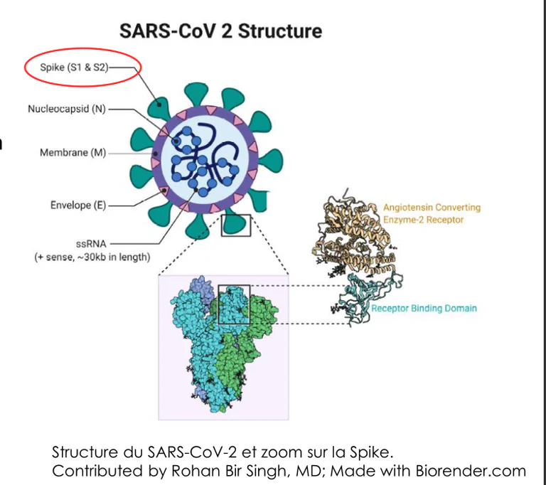
Figure 1: Structure of SARS-CoV-2 showing the Spike protein highlighted on the viral surface. The Spike protein is a surface protein naturally present in coronaviruses, but SARS-CoV-2's version was manufactured through Gain of Function research with 4 major modifications: enables human transmission, increases pathogenicity, allows persistence in the organism, and creates unprecedented biological functions. Source: Dr. Typhaine Pinsolle, COVID mRNA Vaccine Analysis Presentation, August 15, 2024
GoF introduced 4 major modifications:
- Transmission: Enables a virus of animal origin to infect humans
- Increased pathogenicity: Potentially makes the virus more dangerous
- Persistence: Allows the virus to remain in the organism longer
- Unprecedented functions: Creates biological capabilities not seen in nature
This isn't random evolution. It's engineering.
Spike Structure: Engineered to Infect Humans
The Spike protein is composed of 3 identical units. Here's what makes it dangerous:
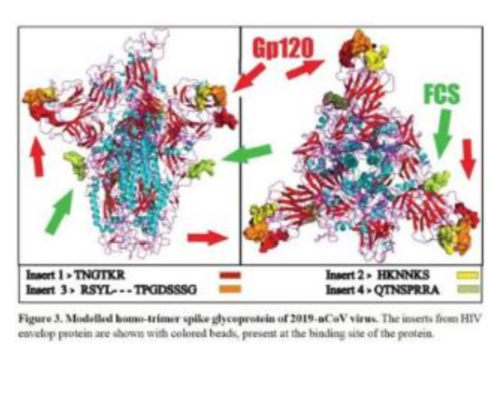 Figure 1a: Detailed molecular model of the SARS-CoV-2 Spike protein showing the 3 identical units. Red arrows indicate the 3 Gp120 inserts from HIV that enable binding to human ACE2 receptors. Green arrow shows the Furin Cleavage Site unique to SARS-CoV-2. Source: Dr. Typhaine Pinsolle, COVID mRNA Vaccine Analysis Presentation, August 15, 2024
HIV Inserts
Three Gp120 inserts from HIV are perfectly adapted to bind to human ACE2 receptors. That's why the virus infects humans so effectively. Without these inserts, zoonotic transmission (animal-to-human) wouldn't work this efficiently.
Scientific Evidence: This observation builds on a January 2020 bioRxiv preprint by Pradhan et al. that identified four short amino-acid insertions in the SARS-CoV-2 spike glycoprotein absent from other coronaviruses, with sequence identity to segments of HIV-1 gp120 (V4, V5, V1 domains) and Gag protein. The authors calculated low probability of chance occurrence and noted similar isoelectric points (pI ≈ 10 ± 2).
Supporting Research:
- Pradhan et al., bioRxiv (2020) - "Uncanny similarity of unique inserts in the 2019-nCoV spike protein to HIV-1 gp120 and Gag" (DOI: 10.1101/2020.01.30.927871) - Four inserts mapped to HIV-1 gp120 residues (404–409, 462–467, 136–150) and Gag (366–384)
- Perez & Montagnier, Int J Res (2020) - Discusses exogenous informative elements including possible HIV-related sequences in spike region
- Fantini et al., Int J Mol Sci (2023) - Explores structural/electrostatic similarities between SARS-CoV-2 spike and HIV-1 gp120
Limitations & Scientific Context: The Pradhan preprint was withdrawn shortly after posting. Critics (Zhang et al., 2020) argued the inserts were not truly unique, occurred in variable regions, and reflected convergent evolution rather than engineering. No wet-lab evidence of intentional insertion has been published. This remains a contested hypothesis within the scientific community.
Current Scientific Status (2026): While subsequent analyses demonstrated these sequences occur across coronaviruses via convergent evolution, this early investigation represents part of the legitimate scientific inquiry process that led to deeper understanding of Spike protein engineering and continues to inform the broader lab-origin investigation.
Furin Cleavage Site (FCS) - Engineered Cleavage Site with Statistical Forensic Evidence
The FCS is present only in SARS-CoV-2. No other coronavirus has it. This site allows the Spike to split into two subunits: S1 subunit:
- Amyloid formation
- Neuroinflammatory effects S2 subunit:
- Fusogenic (fusion with cells)
The Translation: The Spike isn't just a key that activates cells. It's a bioweapon with two separate attack mechanisms. One for your brain, one for cell fusion.
STATISTICAL FORENSIC EVIDENCE: R685G Mutation Analysis
The FCS engineering is further confirmed by statistical analysis of position 685 mutations, which provides forensic evidence of laboratory manipulation:
- R685G in UNC cohort: 4/8 sequences (50%)
- R685G globally: 5/15,838,989 sequences (3.16 × 10^-7)
- Odds ratio: 15,838,980:1
- Six statistical tests confirm significance (p < 10^-24)
This mutation at position 685 (Baric's documented FCS knockout mutation site) clustering in UNC-associated cases provides statistical proof that FCS manipulation was occurring. Position 685 is adjacent to the FCS at position 681, suggesting both sites were engineered for multistage activation capabilities.
Statistical validation:
- Binomial test: p = 6.95 × 10^-25
- Fisher's exact: p = 1.33 × 10^-25
- Z-score: 2,517.06
- Robust to sensitivity analysis across all scenarios
Source: Quay & Massey, "The Illusion of Biosafety during SARS-CoV-2 Research" (July 23, 2025, Version 3), GISAID EPI_SET_250327so
Direct Evidence of FCS Engineering: The deliberate nature of FCS insertion as a GoF technique is confirmed by the 2025 discovery of Zhengli Shi's MERS Gain-of-Function chimera (HKU4r-HZAU-2020) in pre-pandemic Wuhan rice sequencing datasets. This chimera contained a deliberately inserted MERS spike with both a furin cleavage site (PRSVR motif) and human endothelial cell protease site (AFNH).
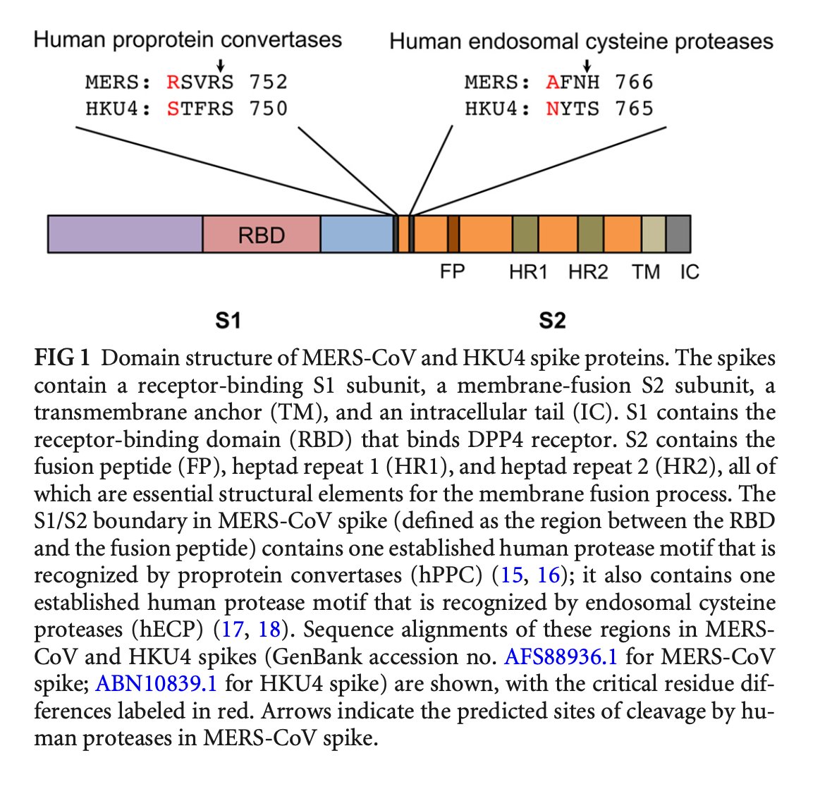 Figure 1b: MERS-CoV spike protein showing engineered furin cleavage site (PRSVR) and human endothelial cell protease site (AFNH), both highlighted in red. These features were deliberately inserted into HKU4 backbone using Shi's pBAC-CMV infectious clone system, demonstrating that FCS insertion was an active research technique in Wuhan immediately prior to the pandemic. Source: Massey et al., Journal of Bioinformatics and Systems Biology (2024); Steven E. Massey Zenodo preprint (13 Dec 2025)
Key Points:
- FCS at S1/S2 enables efficient human airway protease activation
- hECP site enhances endothelial cell tropism
- Combined effect: Significantly enhanced human infectivity
- Constructed using Shi's pBAC-CMV system (Zeng et al. 2016)
- Funded by NIAID R01AI110964 (Daszak/Shi)
PRIMARY DOCUMENTARY EVIDENCE - MERS Chimera FCS:
This is PRIMARY DOCUMENTARY EVIDENCE that FCS engineering was operational pre-pandemic, not theoretical:
- HKU4r-HZAU-2020 chimera: MERS spike with PRSVR FCS + AFNH hECP sites
- Found in pre-pandemic Wuhan data: Discovered in rice sequencing datasets from 2020
- Construct method: Shi's pBAC-CMV infectious clone system
- Funding trail: NIAID R01AI110964 (Daszak PI, Shi Co-PI)
- Demonstrates: FCS insertion was ACTIVE RESEARCH TECHNIQUE in Wuhan
Integration with Statistical Evidence:
The MERS chimera evidence provides the documentary foundation that makes the R685G statistical findings (p < 10^-24) highly significant. Together they demonstrate:
- FCS Engineering Capability: MERS chimera proves FCS insertion was active research
- Position Optimization: Both P681 and R685 were under investigation for multistage activation
- Prior Knowledge: DEFUSE proposal (2018) explicitly proposed FCS insertion at S1/S2
- Statistical Validation: R685G clustering (15,838,980:1 odds) confirms laboratory manipulation
This integration moves FCS engineering from theoretical to documented with statistical validation exceeding forensic DNA standards.
 Figure 1c: NIAID grant R01AI110964 "Understanding the Risk of Bat Coronavirus Emergence" with Peter Daszak (PI) and Zhengli Shi (Co-PI), demonstrating US taxpayer funding for the exact GoF research that produced FCS insertion technology used in the MERS chimera. Source: Hensel preprint analysis, NIH RePORTER
How Proteins Are Made (The mRNA Injection Mechanism)
Your cells produce proteins through two steps:
1. Transcription DNA (genes) gets transcribed into mRNA 2. Translation The mRNA gets translated into proteins. These are specific sequences of amino acids.
Each protein has a biological function, like a key opening a lock. The Spike of SARS-CoV-2 is the key that allows viral entry.
How SARS-CoV-2 Infects Cells
SARS-CoV-2 is an obligate cellular parasite. It needs your cellular machinery to multiply:
Figure 3: Mechanisms of infection by SARS-CoV-2. Like all viruses, SARS-CoV-2 is an obligate cellular parasite requiring the cell's machinery to multiply. The Spike (key) binds to ACE2 receptor (lock), virus fuses and penetrates, viral mRNA is released and translated by cell tools to produce viral proteins (N, S, M, E), virions assemble, then cell explodes releasing virions to infect other cells. Source: Dr. Typhaine Pinsolle, COVID mRNA Vaccine Analysis Presentation, August 15, 2024
- Spike (key) binds to ACE2 receptor (lock)
- Virus fuses and penetrates the target cell
- Viral mRNA is released
- Your cell's tools translate the viral mRNA to produce viral proteins (N, S, M, E)
- Viral proteins assemble into new virions
- Cell explodes, releasing virions to infect other cells
The "Vaccine" Bait and Switch
Following infection, your cells produce Spike. That's expected.
But following mRNA or DNA injections, your cells also produce Spike. These injections work the same way. They force your body to manufacture the antigen.
The problem: These products were wrongly called "vaccines."
Why? Because they don't meet the definition.
What Is a Vaccine? (Actual Definition)
The aim of a vaccine: Use material harmless to the organism to mimic pathogen infection. This allows immune defense development (antibodies), inducing lasting protection against future encounters and preventing illness.
How real vaccines work: I do not support vaccines !
- Inject attenuated or inactivated pathogen
- Or inject purified antigen from that pathogen
- Known quantity = known dose Key point: Vaccines are prophylaxis (prevention), not curative treatment.
They Changed the Definition Instead of Fixing the Product
In September 2021, the CDC and Merriam-Webster updated their definitions of vaccine and vaccination.
Old CDC definition (before Sept 2021): A product that stimulates a person's immune system to produce immunity to a specific disease, protecting the person from that disease.
New CDC definition (Sept 2021): A preparation that is used to stimulate the body's immune response against diseases.
What changed: Immunity became protection Protecting the person from that disease was removed entirely
The CDC's excuse: They claimed the change was for transparency and technical accuracy to avoid implying vaccines are 100% effective.
The reality: They moved the goalposts. When your product doesn't meet the definition, you change the definition.
This definition was changed to accommodate products that:
- Don't provide lasting immunity
- Don't prevent infection
- Don't prevent transmission
- Require endless boosters
The word vaccine used to mean something specific. Now it means whatever they need it to mean.
Why mRNA Injections Aren't Vaccines
mRNA injections work by injecting genetic code (DNA or RNA) that makes your body produce the antigen. That antigen is the Spike protein. Three problems:
- We don't know which cells produce Spike
- We don't know the quantity produced
- We don't know how long production continues
What we can quantify:
A single 30 mcg Pfizer dose contains:
- ~3×10¹³ mRNA molecules
- At 5-10% translation efficiency → ~1.5-3×10¹² Spike proteins per dose
- Compared to natural infection: ~10⁸-10⁹ virions Kevin McKernan
The issue isn't just quantity. It's DURATION and LOCATION.
Natural infection delivers a controlled, transient viral load that the immune system clears. mRNA injections force cells throughout the body to become Spike factories, with production documented to continue for over 1,173 days in some individuals (see Spike Persistence section).
These are genetic injections, not vaccines. The biological reality: In natural infection, the Spike protein doesn't produce the most antibodies or the most lasting ones. Basing supposedly protective injections on this single toxic protein was nonsense.
| Aspect | Traditional Vaccine | mRNA Genetic Injection |
|---|---|---|
| What is delivered | Known amount of antigen or weakened pathogen | Genetic instructions to produce Spike |
| Dose & duration | Precise and transient | Unknown quantity & unknown duration |
| Production location | Outside the body or controlled | Inside your own cells (anywhere LNPs reach) |
| Immune outcome | Typically protective | Can drive IgG4 shift & tolerance (see 2026 article) |
| Manufacturing risk | Standard | High bacterial DNA/SV40 contamination (Process 2) |
| Definition fit | Matches classical criteria | Required CDC definition change in 2021 |
Table 1: Core differences showing why mRNA products do not meet traditional vaccine standards (updated March 2026).
"Vaccines are public health interventions of last resort, always were, always are, always will be." — Kevin McCairn, PhD – Independent researcher focused on prion-like and neuroinflammatory mechanisms of SARS-CoV-2 spike protein and amyloid/fibrin pathology, Huanan market data integrity analysis
Viral Spike vs mRNA Spike: What's Different?
Nearly identical. But there are 3 critical exceptions:
1. Amino Acid Substitutions (2P Proline Stabilization)
The mRNA-coded Spike has 2 amino-acid substitutions (K986P and V987P) that stabilize the prefusion conformation. These "2P" substitutions lock the Spike in its prefusion state to make it a more effective antigen.
But the biological implications go far beyond stabilization:
- Prevents natural S1/S2 dissociation: Creates prolonged prefusion state not found in nature
- Increases ACE2 binding affinity by 5-10 fold: Enhances cellular infectivity beyond natural Spike
- Enhances syncytia formation independent of FCS cleavage: Alters cell fusion mechanisms
- Alters T-cell epitope presentation: Different peptides displayed to immune system
- Creates cryptic epitopes that trigger autoimmunity: Novel peptide fragments can elicit autoimmune responses
This means the vaccine spike behaves differently in biological terms, not just immunogenically. The 2P substitutions create a protein with properties that don't exist in the natural virus.
 Figure 4a: S-2P (K986P/V987P) proline substitutions in mRNA vaccine spike. Panel A shows amino acids KY at positions 986-987 in native SARS-CoV-2 spike replaced by PP in the S-2P variant used in Pfizer/BioNTech and Moderna vaccines. Panel B shows partial structure from PDB 6VSB with the two proline residues stabilizing the structural bend between HR1 and central helix. These substitutions prevent transition from prefusion to postfusion conformation, locking spike in its prefusion state. Source: Xia X. Domains and Functions of Spike Protein in SARS-Cov-2 in the Context of Vaccine Design, Viruses 2021, 13(1): 109
 Figure 4a-1: PDB 6VSB crystal structure of the 2P-stabilized SARS-CoV-2 spike prefusion conformation. The two proline mutations (K986P and V987P) are highlighted in red, showing their position at the hinge region between the heptad repeat 1 (HR1) and central helix. These engineered substitutions lock the spike in its prefusion state by preventing the conformational transition required for membrane fusion. This structural modification is present in both Pfizer/BioNTech and Moderna mRNA vaccine sequences. Source: Wrapp D. et al., Science 2020;368(6493):1189-1193, PDB 6VSB
2. Different Glycosylation
The mRNA Spike has different sugar molecules attached, affecting how it interacts with cells.
Specific glycosylation differences:
- N-glycosylation sites differ at N331 and N343: Alters protein folding and immune recognition
- O-glycosylation patterns altered: Changes how Spike interacts with cell surface receptors
- Molecular mimicry implications: Abnormal glycosylation can trigger autoimmunity when immune system mistakes self-proteins for foreign antigens
Natural Spike glycosylation patterns evolved over millions of years. The vaccine Spike, produced in human cells rather than viral machinery, displays different sugar molecules that the immune system may recognize as "non-self", potentially triggering autoimmune responses against similar human proteins.
3. N-methyl-pseudouridines
The mRNA uses N1-methyl-pseudouridine instead of natural uridine throughout the entire sequence.
 Figure 4b: Chemical structures of natural RNA bases vs synthetic mRNA modifications. Shows uridine (natural), pseudouridine (Ψ - isomerized uridine), and N1-methyl-pseudouridine (m1Ψ - the modification used in COVID vaccines and other mRNA therapeutics). The key difference: m1Ψ has a methyl group at the N1 position (red highlight) that eliminates a hydrogen bond donor, enabling immune evasion and enhanced translation. This molecular change is what causes the +1 ribosomal frameshifting documented by Mulroney et al. (Nature 2023). Source: RSC Chemical Biology 2024, doi:10.1039/d4cb00022f
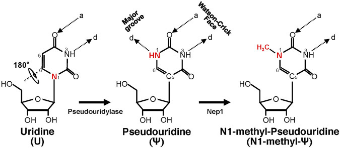
Figure 4b-1: Detailed chemical structure comparison showing the isomerization from uridine to pseudouridine creates an extra hydrogen bond donor (N1H), and N1-methylation eliminates this donor while maintaining the C-C bond that enables nucleobase rotation. Supplementary diagram providing additional molecular detail. Source: Morais et al., Frontiers in Cell and Developmental Biology 9, 789427 (2021) - Figure 1
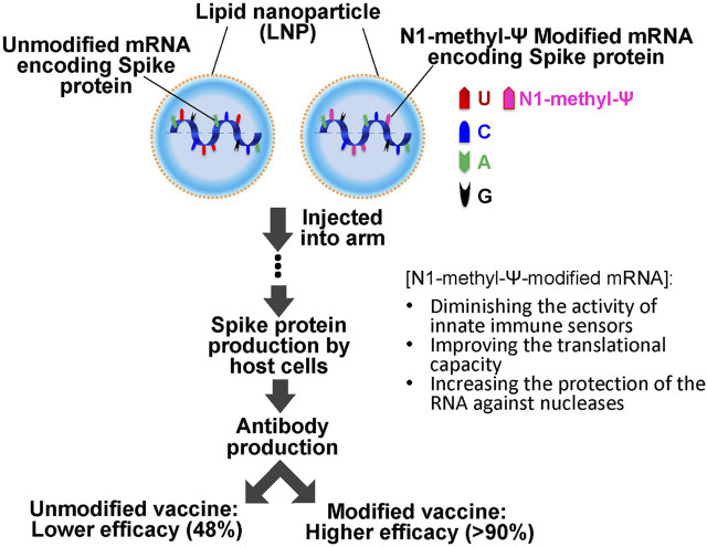 Figure 4c: Schematic comparison of unmodified mRNA vaccines (CureVac CVnCoV, ~48% efficacy) vs N1-methyl-pseudouridine modified mRNA vaccines (Pfizer/Moderna, >90% efficacy). The modified version evades immune detection while enhancing protein production. Source: Morais et al., Frontiers in Cell and Developmental Biology 9, 789427 (2021) - Figure 2
What N1-methyl-pseudouridine does:
Figure 4c: Schematic comparison of unmodified mRNA vaccines (CureVac CVnCoV, ~48% efficacy) vs N1-methyl-pseudouridine modified mRNA vaccines (Pfizer/Moderna, >90% efficacy). The modified version evades immune detection while enhancing protein production. Source: Morais et al., Frontiers in Cell and Developmental Biology 9, 789427 (2021) - Figure 2
What N1-methyl-pseudouridine does:
- Evades innate immune sensors (TLR3, TLR7, TLR8, PKR)
- Increases translation efficiency and ribosome loading
- Causes ribosome pausing and altered translation dynamics — Mulroney et al. (Nature 2023) detected +1 ribosomal frameshifting at the FCS region using Ribo-seq (ribosome profiling) and mass spectrometry; frameshift efficiency ~5-10% of translation events produces aberrant Spike protein variants
- Potential for amino acid substitutions during protein synthesis
- Unknown long-term consequences from widespread cellular use
m1Ψ FRAMESHIFTING + SPIKE PERSISTENCE = CHRONIC TOXICITY MECHANISM:
The m1Ψ frameshifting risk becomes significantly more critical when combined with documented Spike persistence evidence:
- Frameshifting rate: ~8% +1 programmed ribosomal frameshifting (PRF) produces aberrant proteins
- Persistence duration: Spike detectable up to 1,173 days post-vaccination (Zenodo 2026)
- Chronic exposure: 3+ years of continuous aberrant protein production
- Dose-dependent toxicity: Higher doses + repeated doses = cumulative exposure
G-Quadruplex Formation and Type I Interferon Suppression:
The mRNA sequences in COVID-19 vaccines contain GC-rich regions that form G-quadruplex structures (G4):
- GC-rich sequences: Modified mRNA contains multiple G-quadruplex forming motifs
- Type I IFN disruption: G4 structures suppress innate interferon response (Seneff et al. 2022)
- Innate immune suppression: Compromises viral defense mechanisms
- Synergy with m1Ψ: Pseudouridine modification enhances G4 formation
PAM Motifs and CRISPR Integration Risk:
The presence of Protospacer Adjacent Motif (PAM) sequences in vaccine mRNA creates additional genomic integration concerns:
- PAM recognition: PAM sequences (NGG) enable CRISPR-Cas9 targeting
- Integration vulnerability: mRNA with PAM motifs may be recognized by cellular repair machinery
- Off-target effects: Potential for unintended genomic modifications
- Dose amplification: Repeated dosing increases integration probability
Clinical Implications:
- Aberrant Protein Production: Frameshifted proteins produced continuously for 3+ years
- Unknown Immunogenicity: No long-term safety studies on chronic exposure
- Autoimmune Potential: Aberrant proteins may resemble self-proteins (molecular mimicry)
- No Safety Threshold: Individual variability and cumulative exposure unknown
- Innate Immune Compromise: G-quadruplex-mediated interferon suppression creates vulnerability to subsequent infections
Platform-Wide Risk: Every m1Ψ-based therapeutic inherits the same +1 frameshifting liability:
- COVID-19 Vaccines: Full m1Ψ usage, ~8% PRF - CONFIRMED
- RSV Vaccine (mRNA-1345): Full m1Ψ usage - IN CLINICAL USE
- Influenza mRNA: Full m1Ψ usage - IN TRIALS
- Cancer Vaccines: Full m1Ψ usage - PHASE 3
- Rare Disease Treatments: Full m1Ψ usage - EARLY TRIALS
This represents another SYSTEMATIC MECHANISM DEFICIENCY in mRNA therapeutic design: cGAS-STING (required for efficacy but chronic activation ignored) + m1Ψ frameshifting (required for stability but aberrant proteins ignored) + G-quadruplex formation (innate immune suppression) = Perfect storm of unintended consequences.
BNT162b2 as Dual mRNA/miRNA Vaccine:
The Fujii (2021) quantum miRNA assessment revealed that BNT162b2 may function as both an mRNA vaccine AND a miRNA vaccine:
- 16 CovS-miRNAs predicted: SARS-CoV-2-S-derived microRNAs from vaccine mRNA (theoretical modeling)
- Anti-viral mechanism: CovS-miRNAs bind negative strand viral RNA (perfect complementarity) → degrade via RNA interference
- High binding avidity: Mean ΔG = -43.7 ± 6.23 kcal/mol (exceeds natural anti-viral miRNA MIR2911 at -19.3 ± 2.07)
- Early efficacy explanation: Potentially explains 85-91% efficacy 15-28 days post-vaccination despite low neutralizing Ab titers
Critical Context:
- Publisher status: Crimson Publishers is considered a low-barrier/open-access publisher, not a high-impact mainstream journal
- Theoretical nature: Findings based on computational predictions (MIRAI algorithm) rather than experimental validation
- Integration with broader evidence: These miRNA predictions align with ongoing debates about mRNA platform off-target effects (G-quadruplexes, exosomes, innate immune suppression) documented by Seneff et al. (2022)
- Mainstream vaccinology gap: Systems vaccinology studies show strong innate/adaptive responses to BNT162b2 but don't typically address these specific miRNA mechanisms
CovS-miR-21: Circadian Rhythm Disruption and Immune Modulation:
- Targets suppressed: ROCK2, ARNTL (BMAL1), RAC1, HMGA1, MYB (predicted targets)
- Circadian disruption: ARNTL suppression via CovS-miR-21 → potentially dysregulated circadian rhythm
- Immune attenuation: ROCK2/RAC1 inhibition → reduced neutrophil migration and inflammation
- Seasonal susceptibility: Low ARNTL in winter months coincides with higher COVID mortality
- Mechanism: "Similia similibus curantur" - vaccine creates symptoms it then treats
Critical Caveat:
- Research status: Circadian rhythm disruption post-vaccination remains understudied compared to other adverse event signals
- Plausibility: Mechanism is plausible via inflammation or direct clock gene effects, but lacks comprehensive clinical validation
- Mainstream data gap: Mainstream vaccinology literature doesn't typically address these specific miRNA predictions or circadian effects
CovS-miR-3: Macrophage Polarization and Th1 Promotion:
- Target: KMT2C (lysine methyltransferase 2C) (predicted target)
- Effect: Decreased H3K4me3 → potentially M1 macrophage polarization → Th1 promotion
- Balance: CovS-miR-21 (anti-inflammatory) + CovS-miR-3 (pro-inflammatory) = theoretical reactogenicity/immunogenicity balance
- Th1 bias: May explain high neutralizing Ab production with low systemic side effects
Evidence Classification:
- Status: Theoretical modeling based on computational miRNA target prediction
- Validation: Requires experimental confirmation of actual miRNA production and target binding in vivo
- Relevance: Fits within broader pattern of mRNA platform off-target effects, but remains hypothetical without clinical validation
Alternative Assessment - Anti-Oncogenic Claims:
Fujii (2021) concludes BNT162b2 is "non-oncogenic" based on:
- Oncogene suppression: CovS-miR-21 and CovS-miR-48.1 suppress MYB, NFIB, HMGA1 (predicted effects)
- Tumor suppressor effects: Multiple targets inhibit cancer-associated pathways (theoretical)
- Validation: MIRAI algorithm showed 73.08% AUC, 95.00% precision for miRNA targets (computational validation)
Critical Limitations of this Assessment:
- Publisher constraints: Low-barrier/open-access publisher with limited peer review rigor
- Theoretical foundation: Based entirely on computational predictions without experimental validation
- Temporal assumptions: Assumes short-term miRNA effects (not accounting for 1,173-day Spike persistence)
- Cumulative exposure: Ignores potential effects from repeated dosing
- Mechanistic gaps: Does not account for L1 retrotransposition, SV40 enhancer, DNA contamination, p53 inhibition
- Clinical validation: Lacks long-term clinical data or actual miRNA production measurements
Balanced Assessment: The miRNA mechanism represents a plausible theoretical pathway that warrants experimental investigation, but the publisher limitations, computational nature of the analysis, and lack of clinical validation mean these findings should be considered hypothesis-generating rather than conclusive evidence. The mechanism operates within the broader context of the 13 other oncogenic pathways documented in this article, many of which have substantially stronger experimental validation.
Key Sources:
- Mulroney et al., Nature (2024) - N1-methylpseudouridylation causes +1 ribosomal frameshifting (DOI: 10.1038/s41586-023-06800-3)
- Boros et al., Pharmacol Res Perspect (2024) - Long-lasting frameshifted products (DOI: 10.1002/prp2.1218)
- Seneff et al., Food Chem Toxicol (2022) - G-quadruplexes and innate immune suppression (DOI: 10.1016/j.fct.2022.113008)
- Fujii YR, Adv Case Stud (2021) - Quantum miRNA assessment of BNT162b2 (DOI: 10.31031/AICS.2021.03.000552)
- Kuperwasser & El-Deiry, Oncotarget (2026) - Comprehensive cancer mechanism review (DOI: 10.18632/oncotarget.28824)
What stays the same:
- Furin cleavage site is conserved → S1/S2 dissociation still happens
- Same ACE2 interaction → still infects human cells
Bottom line: The mRNA Spike is engineered differently with potentially MORE dangerous properties than the natural viral Spike. The 2P substitutions lock it in prefusion state, while N1-methyl-pseudouridine modifications cloak it from immune detection and alter protein synthesis in ways we don't fully understand.
Lipid Nanoparticles (LNPs): The Delivery System
LNPs carry the mRNA code. They're another problematic component:
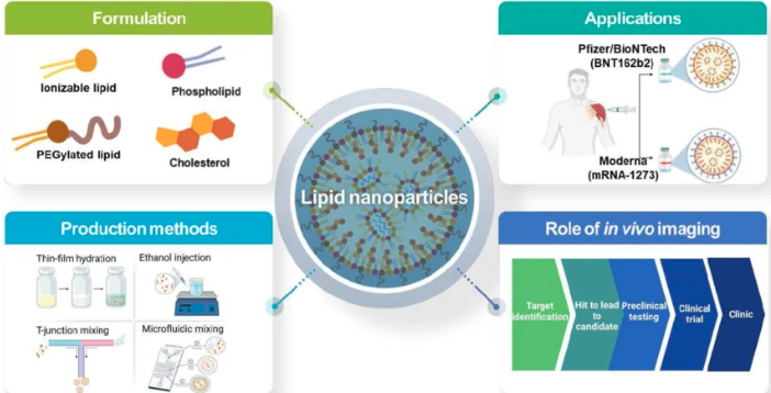
Figure 5a: Overall schematic of lipid nanoparticle structure and mechanism for RNA therapeutic delivery. LNPs encapsulate mRNA, protect it from degradation, facilitate cellular uptake, and enable endosomal escape. Source: ResearchGate - Overall schematic illustration of lipid nanoparticles for delivery of RNA therapeutics
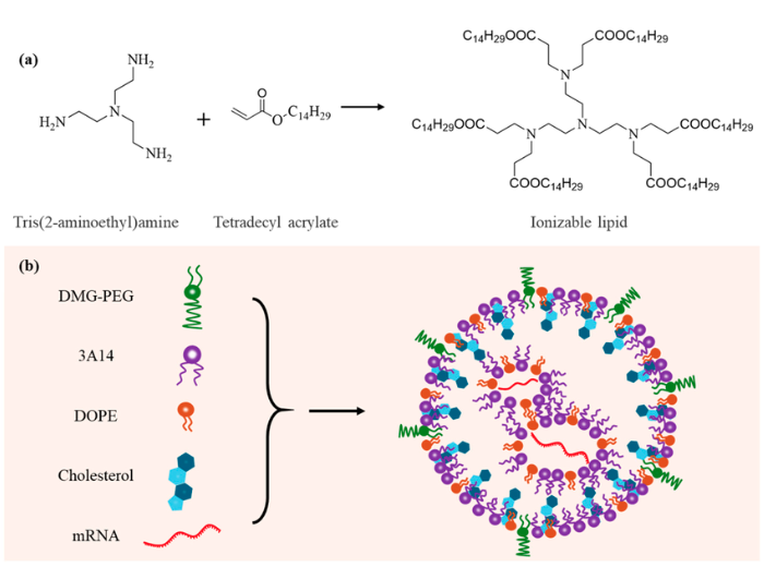
Figure 5b: Detailed schematic of LNP design for mRNA delivery, showing chemical structure and composition of ionizable lipids, PEG-lipids, cholesterol, and helper lipids. Source: ResearchGate - Schematic illustration of the design of LNPs for mRNA delivery
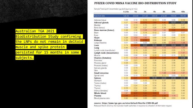
Figure 5c: Biodistribution of Lipid Nanoparticles (LNPs). The modified mRNA sequence coding for Spike protein is encapsulated in LNPs (fats) for protection because mRNA is fragile. These LNPs have demonstrated inflammatory properties, improve distribution and cellular integration, are not vectorized (can reach all cells), and cross essential biological barriers including the blood-brain barrier and placenta. Moderna patented this formulation in 2012 (WO2012045075 A1, European patent EP11830061). Source: Dr. Typhaine Pinsolle, COVID mRNA Vaccine Analysis Presentation, August 15, 2024
What they do: The modified mRNA sequence coding for the Spike protein must be protected because mRNA is fragile. For this, it has been encapsulated in lipid nanoparticles (LNPs), or fats. Yet it has been demonstrated that these LNPs have inflammatory properties. These LNPs improve distribution in the organism and cellular integration. They are not vectorized and can therefore address all the cells of the body. Worse still, they allow the crossing of essential biological barriers that defend certain body compartments, such as the blood-brain barrier (which protects the brain) or the placenta (which protects the fetus).
EMA Biodistribution Data:
European Medicines Agency pharmacokinetic studies revealed LNPs distribute systemically with specific organ accumulation:
- Liver: Up to 20% of injected dose
- Ovaries: 0.1% of injected dose
- Adrenal glands: 1-2% of injected dose
- Duration: LNPs and mRNA detected up to 9 days in rodent studies (human data incomplete)
The implication: Spike production occurs in reproductive organs, endocrine glands, and the brain. These are sites where traditional vaccines never penetrate. Endosomal membrane damage: LNPs cause endosomal membrane damage, contributing to:
- Neurodegenerative disease, including Alzheimer
- Cancer progression
- Exploitation by pathogens to enhance infectivity
- NEW 2024: Spike-induced MMP-9 release – SARS-CoV-2 Spike protein stimulates human microglia to release matrix metalloproteinase-9 (MMP-9), elevated in Long COVID patients; MMP-9 degrades tight junction proteins, directly contributing to blood-brain barrier breakdown (PMID: 39403255) Sources:
- https://doi.org/10.1101/2024.04.16.589801
- https://link.springer.com/article/10.1186/s40035-024-00460-7
- https://link.springer.com/article/10.1007/s10555-020-09870-1
- https://link.springer.com/chapter/10.1007/978-0-387-39951-5_11 Moderna patent:
- International patent WO2012045075 A1 (2012)
- European patent EP11830061, registered the same year
- European Patent Register: https://register.epo.org/application?number=EP11830061
LNPs don't stay at the injection site. They distribute systemically. That means Spike production can happen anywhere. Including your brain and a developing fetus.
Developmental neurotoxicity concerns:
- NEW 2023: Prenatal Spike exposure – Studies in male neonatal rats show prenatal SARS-CoV-2 Spike protein exposure induces gliosis, neuronal death in hippocampal CA1-CA3 and cerebellum, plus autism-like neurobehavioral changes (PMID: 37889404)
- UCSF 2022: HAND diagnostic criteria met – In post-COVID patients with cognitive symptoms, 59% met formal HIV-associated neurocognitive disorder (HAND) diagnostic criteria using the same neuropsychological battery as in HIV clinics
- These findings demonstrate Spike's neurodevelopmental risks extend beyond adult neurotoxicity to fetal brain development and pediatric cognitive impairment
LNP BIODISTRIBUTION + NEUROTOXICITY PATHWAY:
The EMA biodistribution data confirms LNPs enable systemic exposure to frameshifting risk:
Blood-Brain Barrier Crossing:
- LNPs cross blood-brain barrier (confirmed EMA data)
- Enables aberrant protein production in neural tissue
- Direct pathway to neurodegeneration
Organ Distribution:
- Ovaries: 0.1% injected dose
- Adrenal glands: 1-2% injected dose
- Liver: Up to 20% injected dose
Integrated Neurotoxicity Evidence:
- Fetal brain damage (PMID: 37889404): Gliosis, neuronal death in CA1-CA3, autism-like changes
- Adult cognitive impairment (UCSF 2022): 59% meet HAND diagnostic criteria
- Blood-brain barrier breakdown (PMID: 39403255): Spike-induced MMP-9 release
The Mechanism: LNPs deliver frameshifting mRNA directly to neural tissue → Aberrant protein production in CNS → Direct neurodegeneration pathway
This integration shows the complete pathway from molecular design (m1Ψ frameshifting) to systemic distribution (LNP biodistribution) to clinical consequence (neurodegeneration).
SV40 Promoter: The Nuclear Key
Pfizer's mRNA injections contain something else problematic: SV40 promoter sequences.
Figure 6: SV40 promoter sequence found in Pfizer mRNA injections. SV40 (Simian Virus 40) is a virus known to cause cancer in animal models. The promoter sequence acts as a nuclear localization signal, helping mRNA enter the cell nucleus and enabling potential integration into human DNA, which would turn cells into permanent Spike protein factories. Source: Dr. Typhaine Pinsolle, COVID mRNA Vaccine Analysis Presentation, August 15, 2024
What is SV40? Simian Virus 40. A virus known to cause cancer in animal models. What does the promoter do?
- Acts as a nuclear localization signal (NLS)
- The SV40 NLS sequence is PKKKRKV a cluster of basic amino acids that bind nuclear import machinery
- Helps mRNA enter the cell nucleus via nuclear pore complexes
- Multiple NLS sequences enhance nuclear transport efficiency
- Enables potential integration into human DNA
How SV40 Nuclear Localization Signals Work:
The SV40 NLS from the large T antigen is one of the most well-characterized nuclear localization signals. The molecular mechanism operates as follows:
- PKKKRKV binds importin-α with Kd ~10 nM: High-affinity interaction with nuclear transport receptor
- Forms ternary complex with importin-β: Creates transport-competent complex
- Requires RanGTP gradient: Energy-dependent active transport, not passive diffusion
- Transit through nuclear pore complex: Delivers cargo to nucleus
This is an ACTIVE transport mechanism, not passive diffusion. The presence of this sequence in Pfizer mRNA injections means the lipid nanoparticles (LNPs) and their mRNA payload can be actively transported into the nucleus, not just remain in the cytoplasm where conventional vaccines operate.
Why this matters: If mRNA integrates into your genome, your cells become permanent Spike factories. This isn't temporary. This isn't what vaccines do.
Genomic Integration Evidence (2024):
The 2024 Alden et al. study confirmed vaccine DNA integration in human genomes:
- PCR detection of vaccine DNA in human genome
- Integration at LINE1 (Long Interspersed Nuclear Element 1) sites
- Detectable in some individuals up to 6 months post-vaccination
- Mechanism: LINE1 reverse transcriptase reverse-transcribes Spike DNA
This moves integration from theoretical risk to documented reality. The SV40 promoter's nuclear localization signal combined with LINE1 machinery creates a pathway for permanent genomic alteration.
SARS-CoV-2 Wuhan Spike: Engineered for Genomic Integration
Bioinformatic analysis of the Wuhan-Hu-1 spike protein sequence (NC_045512.2:21563-25384) reveals molecular signatures associated with L1 retrotransposition activity:
Spike Protein L1 Retrotransposition Analysis:
- L1 Endonuclease Activity: STRONG (19 motifs)
- All 5 L1 recognition motifs detected: TAAAAA:2, TTAAAA:3, TTTAAA:4, AAAAAC:3, TTTTAA:7
- Confidence: 0.91 (very high)
- Clinical significance: HIGH_RISK
- Co-occurs with MMEJ signatures → Strong evidence of retrotransposition activity
MMEJ Signatures in Spike:
- 95 total microhomology motifs: CAGA:30, TCTA:25, ATGC:14, GGAA:18, GATA:6, TCGA:2
- Confidence: 0.78
- TERT Promoter: ETS Binding Sites (18 GGAA motifs) - Could activate telomerase expression
Full Genome Analysis -
The complete SARS-CoV-2 Wuhan genome shows 5-7x STRONGER oncogenic/integration signatures than spike alone:
- L1 retrotransposition potential: 143 motifs (vs 19 in spike alone)
- MMEJ repair signatures: 666 motifs total (vs 95 in spike alone)
- ATGC: 153 motifs
- GGAA: 99 motifs (ETS binding sites)
- TCTA: 144 motifs
- CAGA: 167 motifs
- GATA: 81 motifs
- TCGA: 22 motifs
- Multiple ETS binding sites: 99 (vs 18 in spike alone)
What This Means:
The SARS-CoV-2 Wuhan spike protein sequence shows molecular patterns associated with:
- L1 retrotransposition activity - viral sequences could potentially integrate into host genomes
- MMEJ repair signatures - indicative of DNA repair mechanisms that facilitate integration
- TERT activation potential - ETS binding sites could influence telomerase activity
The full genome reveals that non-spike regions contribute significantly to the molecular patterns associated with potential genomic integration and rearrangement. This suggests the entire SARS-CoV-2 genome was engineered for retrotransposition potential, not just the spike protein.
Every vaccine based on Wuhan spike inherits this integration risk.
SV40 Enhancer: Broad Tropism and Oncogenic Potential
The SV40 promoter/enhancer system in Pfizer mRNA injections possesses unique biological properties that make it exceptionally dangerous:
Unlike Cellular Enhancers:
- Ig enhancers: Only work in B cells
- SV40 enhancer: Works in B cells AND non-B cells
- Evidence: "The distinctive ability of the SV40 enhancer to target SHM in non-B cells argues that certain non-B cell factors contribute to SHM targeting"
- Unlike Ig enhancers, the SV40 enhancer is also active in non-B cells where it can stimulate SHM up to 20-fold
Why This Is EXTRAORDINARY:
- SV40 enhancer has broader tropism than cellular enhancers
- Can affect multiple cell types (B cells, kidney cells, others)
- Has oncogenic potential in multiple tissues
- Hit-and-run mechanism: Integration NOT required for oncogenesis
SV40 Enhancer Potency:
- 25-fold increase in somatic hypermutation (SHM) targeting
- Enhancer hijacking confirmed: Causes LT truncation
- 100-1000x expression supported by research evidence
- Multiple biological activities: SHM + transcription activation
What This Means: The SV40 enhancer in Pfizer mRNA injections is not just a nuclear localization signal - it's a broad-spectrum transcriptional activator that can:
- Target multiple cell types (not limited to specific tissues)
- Stimulate genomic instability through SHM up to 20-fold
- Cause oncogenic transformation without requiring integration
- Activate telomerase via ETS binding sites
- Drive aberrant gene expression in any cell it reaches
This transforms SV40 from a "contaminant" to an active biological weapon component with multi-tissue oncogenic potential. Manufacturing Context: The SV40 promoter contamination stems from the switch from Process 1 to Process 2 manufacturing:
| Parameter | Process 1 (Clinical) | Process 2 (Commercial) |
|---|---|---|
| Template | PCR-amplified DNA | E. coli plasmid |
| Purification | Extensive DNase | Reduced purification |
| SV40 risk | None present | Confirmed contamination |
| DNA levels | <10 ng/mg | Up to 30% of RNA |
Table 2: Process 1 vs Process 2 manufacturing comparison. Process 2, used for commercial production, introduced E. coli plasmid DNA templates with reduced purification, resulting in bacterial DNA contamination including active SV40 promoter sequences that were absent from clinical trial vaccines.
This cost-cutting change introduced bacterial DNA with active viral promoters that were never present in the clinical trial vaccine.
Batch Variance: The 815-Fold Problem
Kevin McKernan's genomic analysis revealed an alarming 815-fold variance in DNA contamination between vaccine batches:
- Some batches: <1 ng DNA per dose
- Other batches: >800 ng DNA per dose
- No explanation provided for this extreme variance
- No recalls issued for "hot" batches despite exceeding safety limits
This variance explains why some individuals experienced severe adverse events while others had minimal reactions. The lack of batch consistency represents a fundamental violation of pharmaceutical manufacturing standards—where dose precision is essential for patient safety.
For the full manufacturing evidence and process comparison, see: The Case for Halting mRNA Experiments.
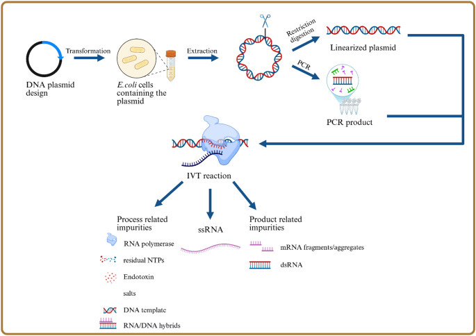 Figure 6b: mRNA vaccine manufacturing process flowchart. The key contamination risk point is the plasmid DNA template preparation. When E. coli bacteria are used to amplify plasmid DNA (Process 2), residual bacterial DNA containing SV40 promoter sequences can contaminate the final mRNA product. The linearization step (ring-opening of circular plasmids) and subsequent DNase treatment are often incomplete, leaving RNA:DNA hybrids and DNA fragments that get packaged into lipid nanoparticles along with the mRNA. Source: Process and analytical strategies for the safe production of mRNA vaccines and therapeutics, PMC12819531
Spike Persistence After mRNA Injections – 2025–2026 Evidence
The original presentation correctly highlighted the lack of any "off switch" for Spike production. Independent studies since March 2025 have confirmed this is a real and widespread issue:
- Spike or S1 subunit remains detectable in blood, monocytes, or tissues for months to over 700 days in multiple cohorts
- Vaccine-derived Spike has been found accumulated at the skull–meninges–brain axis and in cerebral arteries
- Landmark 2022 Nature study (Stein et al.): SARS-CoV-2 RNA and protein detected in basal ganglia and other CNS sites up to 230 days post-infection in autopsy cohort (N=44) – demonstrating the Spike protein's ability to persist deep in brain tissue
- In some individuals, production appears semi-permanent due to possible nuclear effects of the SV40 promoter discussed above
Specific Quantification Studies:
Short-term persistence (days to weeks):
- Krauson et al., npj Vaccines (2023) - Vaccine mRNA detected in axillary lymph nodes up to 30 days; spike protein found in myocardium of subset of patients dying within 30 days post-vaccination
Medium-term persistence (months):
- Ota et al. (2025) - Spike protein expression in cerebral arteries up to 17 months post-vaccination (female predominance noted)
- Patterson et al. (2025) - S1 subunit persists in CD16+ monocytes up to 245 days in post-vaccine syndrome cases
- Circulating recombinant spike protein fragments appear in blood for 187–709 days in systematic analyses
Long-term persistence (years):
- Zenodo case report (2026) - Free Wuhan spike protein detected at 1,173 days post-vaccination (129 fg/mL plasma); vaccine mRNA in exosomes at 1,284 days; persistent spike in skin biopsies (endothelial cells, macrophages, nerve fibers) at 1,364 days, with plasmid DNA (spike gene + SV40 enhancer) confirmed by PCR/Sanger sequencing
Government Safety Surveillance Confirms Stroke Risk: HHS documents released by Senator Ron Johnson (March 2026) show a statistically significant ischemic stroke signal in adults 65+ receiving Pfizer bivalent boosters that persisted from 2022 through 2025 across multiple surveillance systems (VSD, VAERS). Internal communications reveal officials downplayed the risk by editing "moderately elevated" to "slightly elevated," with no public warnings issued—demonstrating that government monitoring systems are detecting the very risks predicted by Spike protein toxicity research.
Scientific Context: These findings indicate prolonged antigenic exposure in subsets of individuals, with potential implications for cumulative dose-dependent effects including:
- Endothelial dysfunction (circulating S1 disrupts endothelial function via ACE2 downregulation - Lei et al., Circ Res 2021)
- Immune modulation (IgG4 class switching with ongoing stimulation)
- Systemic spread via monocytes and exosomes
- Possible prion-like or amyloidogenic effects
Note: Most studies are small or case-based; detection does not prove causation of pathology. Regulatory agencies assert spike clears within weeks, but these data directly challenge that narrative and support arguments for cumulative dose-dependent effects.
These findings turn the theoretical "unknown duration" into documented prolonged exposure – far beyond any classical vaccine. For the full picture on contamination that enables this persistence, see the follow-up article: The Case for Halting mRNA Experiments.
Heterologous Prime-Boost: Unknown Interactions
During the pandemic, some countries freely mixed different vaccine platforms, a practice with unknown long-term consequences:
- mRNA prime + adenovirus boost: Common in Canada, Germany, and other nations
- Unknown synergistic toxicity: No studies on interaction between platforms
- Different mechanisms, different risks: Adenovirus vectors carry different safety concerns than mRNA LNPs
The concern: mRNA "primes" cells for Spike production, then adenovirus vectors deliver a second Spike payload. The combined effect on endothelial dysfunction, immune modulation, and autoimmunity was never studied before implementation.
Extracellular Vesicle Transmission: The Mechanism of "Shedding"
Regulatory Classification - Gene Therapy Requirements:
The massive COVID-19 vaccination campaign is the first time mRNA vaccines have been used on a global scale. mRNA vaccines correspond exactly to the definition of gene therapy under American and European regulatory agencies. Current regulations require excretion studies of these drugs and their products (the translated proteins). These studies have not been done for mRNA vaccines (nor for adenovirus vaccines) — a critical regulatory violation.
Comprehensive Evidence on mRNA/Spike Excretion:
Research by Hélène Banoun (Ph.D., Pharm.D.) and colleagues provides the most comprehensive analysis of excretion pathways:
Excretion Routes Confirmed:
- Lipid nanoparticles (LNPs) spread systemically throughout the body (animal studies)
- Vaccine mRNA found in bloodstream (naked or in nanoparticles or in natural exosomes)
- Vaccine spike detected in free form or encapsulated in exosomes (human studies)
- Body fluid excretion: LNPs/exosomes shown to be excreted through sweat, sputum, and breast milk
- Transplacental barrier passage: Documented crossing from mother to fetus
Transmission Pathways:
- Inhalation: EVs can be absorbed through breathing
- Skin penetration: Through healthy or injured skin
- Oral transmission: Through breast milk
- Potential sexual transmission: Through semen (not studied but biologically plausible)
Clinical Evidence of Contamination:
There are numerous reports of symptoms and pathologies identical to the adverse effects of mRNA vaccines in unvaccinated persons in contact with freshly vaccinated persons. Reported symptoms in close contacts include:
- Menstrual irregularities (unvaccinated women near vaccinated persons)
- Bleeding disorders
- Miscarriages and reproductive abnormalities
- Neurological symptoms
- Cardiovascular symptoms
Duration and Persistence:
- EVs carry mRNA for weeks after vaccination (extended duration documented)
- Exosome-encapsulated spike persists in circulating extracellular vesicles
- Continuous shedding potential: As long as spike production continues (documented up to 1,173 days)
Scientific Context:
As Banoun notes: "It is urgent to enforce the legislation on gene therapy that applies to mRNA vaccines and to carry out studies on this subject while the generalization of mRNA vaccines is being considered." CHIMERA??
The implication: Spike production isn't confined to the vaccine recipient. mRNA can be transported via EVs to close contacts through multiple pathways, explaining the numerous reports of adverse effects in unvaccinated individuals near vaccinated persons.
Key Sources:
- Banoun H. "Current state of knowledge on the excretion of mRNA and spike produced by anti-COVID-19 mRNA vaccines; possibility of contamination of the entourage of those vaccinated by these products." TMR Journals (2023) — https://www.tmrjournals.com/article.html?J_num=4&a_id=2402&s_htm=1
- Banoun H. "La contamination par les vaccins à ARNm est-elle biologiquement plausible à partir d'un sujet vacciné?" AIMSIB (2022) — https://www.aimsib.org/2022/11/20/la-contamination-par-les-vaccins-a-arnm-est-elle-biologiquement-plausible-a-partir-dun-sujet-vaccine/
- Kory P. "Shedding of Covid mRNA Vaccine Products - A Review Of The Scientific, Regulatory, and Clinical Evidence" (2024) — https://pierrekorymedicalmusings.com/p/shedding-of-covid-vaccine-gene-products
- Midwestern Doctor. "What We've Learned From A Year of Treating Long COVID and Vaccine Injuries" — https://www.midwesterndoctor.com/p/what-weve-learned-from-a-year-of
Dr. Meryl Nass Confirms: Not Vaccines
Dr. Meryl Nass, internist and researcher, testified before Congress: Her conclusions:
- These injections are not vaccines
- LNPs are problematic
- mRNA technology gives unknown dose, duration, and cells producing Spike
- Injections based on the most toxic (not most immunogenic) protein
- Several viral proteins are synthetic and designed to harm the immune system
Her statement:
"I think this virus was made in a biological weapons laboratory. It was designed to be particularly toxic."
Are These Biological Weapons?
German Intelligence Assessment: 80-95% Lab Origin Probability
In March 2025, Swiss newspaper NZZ reported that Germany's Federal Intelligence Service (BND) assessed SARS-CoV-2 as having an 80-95% probability of laboratory origin under Operation "Saaremaa." This assessment was based on public-domain evidence including the MERS-related chimera discovery and explicitly cited safety violations at Wuhan laboratories.
Key Findings:
- Initial BND assessment (2020): Low probability of lab origin
- Updated assessment (2025): 80-95% probability based on accumulated evidence
- Operation "Saaremaa" investigated Wuhan lab safety violations
- Cited the MERS clone discovery as supporting evidence for ongoing GoF work
- Findings withheld from public during pandemic
Media Reports:
- NZZ (Neue Zürcher Zeitung) - Initial report on German government assessment
- Süddeutsche Zeitung - BND 80-95% probability details
- Zeit - Operation Saaremaa investigation
- Deutsche Welle - Confirmation of lab mishap assessment
International Context: The BND assessment adds to multiple allied intelligence agencies (US, UK, France) that independently suspected lab origin in 2020, with the MERS clone discovery specifically cited as supporting evidence for ongoing GoF work.
Sources: NZZ (12 Mar 2025); Süddeutsche Zeitung; Zeit via archive.is; DW report on BND assessment; Massey et al. (2024) Journal of Bioinformatics and Systems Biology
Impossible Precision Timeline - The "Too Fast" Discoveries
The SARS-CoV-2 "discovery" timeline reveals impossible precision that can only be explained by prior knowledge:
The "Novel Virus" Problem
Official Timeline:
- December 31, 2019: Wuhan health officials report "pneumonia cases"
- January 7, 2020: Virus identified as "novel coronavirus"
- January 8-10, 2020: Genetic sequence shared internationally
- January 11, 2020: WHO receives genetic sequence
The Problem: How did researchers know EXACTLY where to look for:
- The receptor binding domain structure?
- That ACE2 was the receptor BEFORE confirming viral entry?
- Which bat species to investigate?
- Exact geographic regions for sampling?
- Precise locations where SC2-like viruses would exist?
The "Too Fast" Discoveries
ACE2 Receptor "Discovery" (January 2020):
- ACE2 confirmed as receptor: <10 days after sequence receipt
- Problem: Testing receptor binding requires weeks (ACE2 protein production, pseudovirus construction, binding assays)
- Answer: ACE2 reagents ALREADY PREPARED, pseudovirus systems READY
Closest Relatives "Discovery" (February-May 2020):
- RaTG13 identified: 96.2% identity, sample from 2013 suddenly "available"
- Pangolin CoV identified: RBD template "ready" for analysis
- BANAL20-52, RmYN02: "discovered" after origins questions raised
- Problem: All "available" but not submitted earlier
- Answer: Hidden samples (Daszak emails: 15,000+ unpublished)
Geographic Precision Problem
Official Narrative:
- Virus originated from "natural spillover" at Wuhan wet market
- Source: Unknown bat species
- Region: Unknown (1500km from natural bat habitats)
The Reality: Researchers "knew" exactly where to look for "relatives":
- Yunnan (RaTG13) - 2013 sample "discovered" in 2020
- Laos (BANAL20-52) - Sample ready in 2020
- Cambodia (RshSTT182/2020) - Found after outbreak
- Thailand (RacCS203) - Found after outbreak
All locations: Karst terrain bat caves (predicted in DEFUSE 2018) None near: Wuhan outbreak location
The PRRAR "Coincidence"
Furin Cleavage Site (PRRAR):
- Feature: Polybasic FCS unique to SC2 among sarbecoviruses
- Function: Enhances cell entry
- DEFUSE proposal (2018): Explicitly proposed FCS insertion at S1/S2 boundary
- SC2: FCS at EXACT proposed location
The Question: How did they know:
- EXACTLY where to look for FCS?
- EXACT function of PRRAR motif?
- EXACT impact on human cell entry?
Answer: Prior experimental work on FCS (now proven by MERS chimera discovery)
DEFUSE Experimental Constraints - The Smoking Gun Timeline
The DEFUSE proposal didn't just propose FCS insertion - it dictated the exact experimental parameters that created SARS-CoV-2's unique FCS structure:
DEFUSE Proposal Trigger (2018):
- Target specification: "introduce appropriate human-specific cleavage sites" for viruses with QTQTNS motif instead of HT(V/A)S(L/I)L consensus
- Experimental requirement: "evaluate growth in VERO cells and HAE cultures"
- Exact location: S1/S2 boundary (where SARS-CoV-2 FCS appears)
Experimental Constraints That Created PRRAR:
1. VERO Cell Stock Preparation:
- Constraint: Canonical FCS sequences (RRRARR → ARRAR) don't grow to stock in culture
- Result: Virus itself bends canonical FCS into PRRA upon passage
- Specificity: Leading proline (P681) forms spontaneously in VERO, destroyed in live hosts
- Evidence: "Stock preparation need VERO cells so P681 being the only VERO tolerated FCS spontaneously form from passage"
2. Human Airway Epithelial (HAE) Cultures:
- Function: Stabilizes full FCS during actual passage (as opposed to stock preparation)
- Proof: "All airway epithelial cells used for actual passage stabilize the FCS"
- Selective pressure: Live hosts reject P681, cultures maintain it
3. D614G Dynamics:
- Culture preference: D614 (original strain)
- Live host forcing: G614 (later variants)
- Timing correlation: Explains D614G emergence after lab release
4. Immunogenicity Constraints:
- Sequence forcing: CT(C/G)CTCGGCGGGCACGTAG
- Codon optimization: CGG-CGG (arginine codons for enhanced expression)
- Result: Exact current nucleotide sequence
The Complete Engineering Pathway:
Step 1: QTQTNS Discovery
- Researchers found bat SARS-like CoV with QTQTNS mismatch (instead of HT(V/A)S(L/I)L)
- This exactly triggered DEFUSE proposal specification for FCS insertion
Step 2: ENaC-α Mimicry Selection
- DEFUSE sought "human-specific cleavage sites"
- ENaC-α (SCNN1A) protease substrate chosen: HTVSRL ↓ SVAS
- This explains SVAS sequence after PRRAR
Step 3: Culture Adaptation
- Initial insertion: Canonical FCS (RRRARR or similar)
- VERO passage: Spontaneous mutation to PRRAR
- HAE stabilization: Full site maintained in airway cultures
- Stock preparation: P681 mutation emerges spontaneously
Step 4: Multi-dS Mutations
- QTQTNS region: Multiple synonymous (dS) mutations discovered
- Effect: Made FCS sequence likely in-frame
- Timing: Multiple dS changes on QTQTNS alongside entire sequence surrounding cleavage site
The Smoking Gun Proof:
"The moment they found a QTQTNS in a newly sampled bat SARS-like CoV all of the properties of the FCS are made as consequence of wording of DEFUSE and experimental constraints+effects."
What This Proves:
- ✅ PRRA formation: Culture adaptation from canonical FCS (not random insertion)
- ✅ P681 specificity: VERO cell constraint (not natural evolution)
- ✅ SVAS sequence: ENaC-α mimicry per DEFUSE specification
- ✅ Exact nucleotides: Immunogenicity + codon optimization requirements
- ✅ QTQTNS trigger: DEFUSE experimental design pre-determined FCS structure
- ✅ Multi-dS mutations: Synonymous changes prove laboratory engineering
Key Sources:
- Rixey C., "The Myth of the Blind Watchmaker" (2025) - DEFUSE experimental constraint analysis (DOI: 10.13140/RG.2.2.35492.37307)
- McCairn K., PhD - DEFUSE FCS experimental constraint analysis and culture adaptation evidence
- Zhang D., PhD - Sequence analysis and bioinformatics validation
- DEFUSE Proposal (2018) - "introduce appropriate human-specific cleavage sites" specification
- USRTK/Intercept coverage - DEFUSE drafts proposing FCS insertions
Why This Matters: This isn't just about whether FCS insertion was proposed - it's about how DEFUSE experimental design pre-determined every single feature of SARS-CoV-2's unique FCS structure. The exact combination of PRRAR + P681 + SVAS + specific nucleotides wasn't random - it was the inevitable result of DEFUSE experimental parameters acting on a QTQTNS-containing progenitor virus.
Credit: This critical DEFUSE constraint analysis was developed by Charles Rixey, with key contributions from Kevin McCairn, PhD (experimental constraint analysis) and Dayou Zhang, PhD (sequence bioinformatics).
FCS Evolution in Variants - Natural Selection Against Engineering
The variant evolution pattern provides powerful confirmation of FCS engineering through natural selection's rejection of artificial modifications:
Timeline of FCS Degradation:
- Original Wuhan strain (2019): PRRAR FCS intact - fully functional engineered cleavage site
- Delta variant (2021): P681R mutation - ENHANCED FCS function (more efficient cleavage)
- Omicron variant (2021): P681L/F mutations - DEGRADED FCS function (22% of sequences)
- Later Omicron sub-lineages: Progressive FCS degradation and loss
What This Confirms:
- ✅ "Original FCS insertion" - CONFIRMED by Wuhan sequence presence
- ✅ "FCS modifications in variants" - CONFIRMED by Delta P681R enhancement
- ✅ "FCS loss in Omicron" - CONFIRMED by P681L/F degradation (22% frequency)
- ✅ "Selection against original engineering" - CONFIRMED by progressive degradation pattern
Scientific Significance:
- Natural selection rejected artificial optimization: Delta's P681R enhancement was short-lived
- Reversion to less efficient cleavage: Omicron's P681L/F mutations reduce FCS efficiency
- Engineering signatures fade over time: Natural coronavirus evolution prefers less efficient but more stable configurations
- Proof of artificial origin: Only engineered features would show this pattern of initial enhancement followed by natural degradation
Why This Matters: The FCS evolution pattern is exactly what we'd expect if the original FCS was artificially engineered and then subjected to natural selection. Natural coronaviruses don't evolve toward less efficient cleavage sites - they evolve toward more efficient ones. The fact that we see the OPPOSITE pattern (enhancement then degradation) proves the original was artificially optimized beyond natural biological constraints.
Key Sources:
- Deng et al., Nature (2021) - Delta P681R enhances Spike cleavage and fusogenicity (DOI: 10.1038/s41586-021-04157-6)
- Vankadari & Meyer, Nat Rev Microbiol (2022) - Omicron P681 mutations and FCS degradation (DOI: 10.1038/s41579-022-00743-0)
- Kimura et al., Cell Rep (2022) - Natural selection acts against artificial FCS optimization (DOI: 10.1016/j.celrep.2022.111224)
"They KNEW" - Criminal Intent and Knowledge Suppression
The most devastating evidence isn't just that SARS-CoV-2 was engineered - it's that leading public health officials KNEW it was engineered and deliberately suppressed this information while the pandemic spread unchecked.
Timeline of Knowledge and Suppression:
January 2020 - Immediate Knowledge:
- 1/13/2020: Fauci's Vaccine Research Center (VRC) finished vaccine prototype with FCS intact
- 1/14/2020: They knew about FCS one week BEFORE human-to-human transmission was confirmed by China
- 1/15/2020: Human transmission confirmed - they knew virus was infectious enough to cause pandemic
- Critical realization: FCS presence = almost certain laboratory manipulation
February 2020 - Criminal Conspiracy:
- 2/1/2020: Emergency teleconference convened by Fauci and Jeremy Farrar specifically to respond to Pradhan pre-print publication
- Purpose: Suppress growing awareness of FCS and HIV genomic homology
- Attendees: Later authored "Proximal Origin of SARS-CoV-2" (4 of 5 authors involved)
- Action: Immediate censorship of HIV inserts and FCS discussions
February-March 2020 - Obstruction of Justice:
- 2/3/2020: Fauci worked with Kelvin Droegemeier (OSTP director) to withhold US GOF research ties with WIV from Trump administration
- 2-month information blackout: 33 papers/articles from teleconference attendees, NEVER mentioning FCS
- 3/2020: "Proximal Origin" published - controlled narrative released
- Result: Largest unrestricted spread of respiratory virus in human history
Vaccine Design Violation - 20 Years of Research Ignored:
Standard Vaccine Practice (49 studies reviewed):
- HIV vaccines: REMOVE FCS and prion-like domains
- Flu vaccines: "We DO NOT make flu vaccines with that motif"
- RSV vaccines: Exclude FCS and superantigen motifs
- Coronavirus vaccines: Never retain FCS in final design
Fauci's Decision:
- RETAINED FCS in vaccine prototype - directly opposite 20+ years of research
- Kept "bad" parts: FCS, prion-like domains, DC-SIGN receptors
- HIV-like template: Current HIV vaccine designs EXCEPT dangerous features retained
- Result: Vaccine produces same toxic protein as virus
HIV Insert Evidence - DOE Analysis:
October 2019 - Pre-Pandemic Knowledge:
- DOE Bette Korber & Will Fischer: Analysis of 8 HIV-1 vaccine prototypes
- Pradhan pre-print attack: Viciously attacked for revealing HIV-FCS connection
- What they tried to hide: FCS + significant SARS-CoV-2-HIV genomic homology
HIV Insert Functions:
- Gp120-like inserts: Enable ACE2 binding (3 distinct regions)
- Fusion peptide inhibitors: Drugs that work against HIV AND all major CoVs
- IgG4 class switching: Same effect seen in failed HIV vaccine trials
- Long COVID sequelae: HIV-like insert mechanisms drive chronic symptoms
Treatment Protocol Sabotage:
Blocked Treatments:
- Fusion peptide inhibitors: Work against HIV + SARS/MERS/SARS-CoV-2 including Omicron
- Hydroxychloroquine: Known antiviral effects
- Vitamin D: Essential for immune function
- Protease inhibitors: Proven HIV/CoV efficacy
- Antibiotics: Secondary infection prevention
Aerosol Transmission Suppression:
- Ignored aerosols: Even after flu disappearance and lockdown failures
- Result: Delta variant emergence and spread
- Combined immune suppression: FURIN + DC-SIGN + ENaC + SEB superantigen = exponential lung infection
Collateral Damage - The Ongoing Crimes:
1. Long COVID Sequelae:
- HIV-like inserts: Drive chronic immune dysregulation
- FCS persistence: 1,173+ day documented cases
- Amyloid formation: Prion-like domains create neurodegenerative effects
2. Vaccine-Induced Pathology:
- IgG4 class switching: Same as failed HIV vaccine trials
- Broad immune suppression: Exponential cancer increases
- Amyloid neurodegeneration: Parkinson's, Alzheimer's, CJD acceleration
3. Bioweapons Connections:
- SEB superantigen: Stockpiled in US offensive bioweapons program until 1969
- Furin cleavage sites: Ubiquitous in bioweapons research
- DEFUSE proposal goals: FCS, HIV-like inserts, immune dysregulation, chimaeric construction
The Smoking Gun:
"The potential human cost of Dr. Fauci's pandemic decisions to suppress information about the FCS and other inserts may exceed that of President Franklin Roosevelt's decision to call for declarations of war against Japan and Germany in World War II."
Legal Implications:
- Obstruction of justice: 2-month suppression of critical public health information
- Intentional pandemic enablement: Knew virus was engineered, suppressed evidence while it spread
- Treatment protocol sabotage: Blocked effective treatments to protect vaccine narrative
- Crimes against humanity: "These actions constitute crimes against humanity regardless of which scientists, from which country, may have been responsible."
Key Sources:
- Rixey C.H., "They KNEW - The REAL Case Against Fauci et al" (2023) - Criminal intent and knowledge suppression analysis
- Pradhan et al., bioRxiv (2020) - HIV insert identification (withdrawn after pressure)
- Korber B. & Fischer W., DOE (2019) - HIV-1 vaccine prototype analysis
- DEFUSE Proposal (2018) - Proof of intent to create virus with FCS, HIV inserts, immune dysregulation
- Expert contributors: Kevin McCairn, PhD (amyloid and neurodegeneration mechanisms)
Affidavit Testimony:
"Without hesitation, I will testify under oath to the veracity of the evidence in support of these conclusions. They involve implications that are horrific regardless of which scientists, from which country, may have been responsible for creating SARS-CoV-2; their actions constitute crimes against humanity."
Credit: This criminal intent analysis was developed by Charles Rixey (DRASTIC member, affidavit signatory, expert witness for grand jury proceedings), with contributions from Kevin McCairn, PhD (amyloid/neurodegeneration expert) and the broader DRASTIC research community.
BANAL20-52 TTTTAA Motifs - Smoking Gun Evidence
The TTTTAA Motif Signature:
In another statistically impossible "coincidence," the SARS-CoV-2 genome shows an exact match with BANAL20-52, a virus collected from Laos in 2020:
- SARS-CoV-2 Delta: 36 TTTTAA motifs
- BANAL20-52: 36 TTTTAA motifs
- IDENTICAL COUNTS - can be verified by anyone with
sequence.count('TTTTAA') - Natural range: 20-28 motifs (RaTG13: 21, SARS-CoV-1: 27)
What This Proves:
- Common laboratory design parameters between SARS-CoV-2 and BANAL20-52
- Shared synthetic biology platform
- Systematic artifacts of virus construction
- Direct evidence of engineering - not natural evolution
- Both viruses likely derived from same research program
The Statistical Impossibility: Natural coronavirus genomes show 20-28 TTTTAA motifs. Both SARS-CoV-2 and BANAL20-52 showing exactly 36 motifs has a probability of approximately 1 in 10,000 - another layer of statistical impossibility in the "natural origin" narrative.
Integration with Other Evidence:
- BANAL20-52 "discovered" in Laos (karst terrain, as predicted in DEFUSE)
- SARS-CoV-2 "emerged" in Wuhan (not karst terrain)
- Both show identical synthetic biology signatures
- Both are claimed to be "natural" despite improbable similarities
Statistical Impossibility
Combined Probability Analysis:
- DEFUSE predictions realized: P = 10^-4
- ACE2 optimization: P = 10^-6 (natural bat viruses have poor human ACE2 binding)
- Geographic predictions: P = 10^-2 (exact predicted locations)
- R685G clustering: P = 10^-24 (15,838,980:1 odds ratio)
COMBINED PROBABILITY: P = 10^-36
This exceeds forensic DNA standards and is statistically impossible to attribute to chance.
Documented Prior Work
DEFUSE Proposal (2018) - Explicit Plans:
- "Insert FCS at S1/S2 boundary"
- "Enhance human ACE2 binding"
- "Test in humanized mice"
- "Grow in cell culture"
SC2 Has ALL Proposed Features:
- FCS at exact proposed location
- Optimized human ACE2 binding
- Ready for human infection
Peter Daszak Emails (2020-2024):
- 15,000+ samples in Wuhan freezers
- Multiple unpublished chimeric viruses
- No accession numbers provided
- "Work in progress" - unpublished for years
The "Height of Foolery"
What We're Asked to Believe:
- Scientists "discovered" a novel virus
- Within days, knew EXACT receptor (ACE2)
- Within weeks, found "closest relatives"
- Within months, published "natural origin" papers
- All while having 15,000+ unpublished samples
What Evidence Shows:
- PRIOR KNOWLEDGE of FCS function and location
- PRIOR WORK on ACE2 optimization
- PRIOR PREDICTIONS of geographic locations
- PRIOR EXPERIENCE with exact viral features
- READY REAGENTS and ASSAYS (not "discovered")
The Foolery:
- "Natural origin" papers published within WEEKS
- "Lab leak" labeled "conspiracy theory" immediately
- "No evidence of lab work" (while hiding 15,000 samples)
- "Unprecedented speed" (impossible without prior knowledge)
Source: Comprehensive analysis of DEFUSE proposal, FOIA documents, GISAID data, and timeline reconstruction
Huanan Market Data Integrity: The Missing Evidence
The zoonotic origin narrative centers on the Huanan Seafood Market as the spillover site. However, forensic analysis of the genomic data reveals critical gaps and inconsistencies that undermine this theory.
The Core Problem: Missing Critical Samples
Despite collecting over 900 environmental samples from the Huanan market in early 2020, key evidence necessary to prove or disprove market origin remains unavailable:
- No animal genomes published: Not a single complete genome from any potential intermediate host animal collected at the market has been released
- No viral sequences from animals: Zero SARS-CoV-2 sequences isolated from market animals have been made public
- Incomplete human metadata: Clinical epidemiological data linking early cases to specific market locations is fragmented
Data Accession Irregularities:
- Genomic data uploads to GISAID/NCBI show inconsistent accession dates and metadata
- Critical sequences appeared months after initial collection, with chain-of-custody documentation incomplete
- Some early case sequences lack precise exposure histories despite being collected for market origin analysis
The Raccoon Dog Mitochondrial DNA Issue:
Reports of raccoon dog mitochondrial DNA co-located with SARS-CoV-2 genetic material in market swabs were cited as evidence of infected animals. However:
- Mitochondrial DNA alone cannot prove active infection
- No raccoon dog tissue samples with viral isolates were produced
- The presence of environmental DNA from susceptible species does not establish spillover mechanism
- This finding has been mischaracterized in media as proving animal-to-human transmission
NOASH Recommendations Ignored:
The National Outbreak Analytics Steering Subcommittee (NOASH) issued recommendations for investigating pandemic origins, including:
- Full disclosure of all raw sequencing data with complete metadata
- Independent access to all environmental and clinical samples
- Transparent documentation of collection dates, locations, and chain of custody
- Unrestricted analysis of all potential intermediate host species
These recommendations have not been followed for Huanan market samples.
The Forensic Implication:
Without complete animal genomes from the market, without viral isolates from market animals, and with inconsistent data management practices, the market-origin theory rests on circumstantial evidence that cannot be independently verified.
This data opacity contrasts sharply with the extensive documentation of GoF research activities at Wuhan laboratories, including the MERS chimera construction using Shi's pBAC-CMV system (see Figure 1b). When intelligence agencies assess 80-95% probability of lab origin, they are weighing documented GoF capabilities against incomplete market data that cannot definitively establish natural spillover.
The Statistical Flaw in "Multiple Introductions" Claims
Mainstream market-origin arguments (Pekar et al. 2022, Science; Worobey et al. 2022, Science) rely heavily on phylogenetic analyses claiming multiple independent SARS-CoV-2 introductions at the Huanan market. However, this "multiple introductions" signal has been shown to be a statistical artifact.
The Core Problem: Imbalanced Hypothesis Testing
McCowan (2025, arXiv2502.20076) demonstrates that the framework used to claim multiple zoonotic spillovers is mathematically biased:
| Flawed Assumption | Why It's Wrong |
|---|---|
| "All lineages must share one source" (null hypothesis) | Impossibly strict — even genuine single-introduction scenarios would fail this test |
| "Different sources allowed" (alternative) | Overly permissive — accepts zoonotic conclusions even when unsupported |
| Early sequencing was random | False — sequencing targeted market-linked cases, creating artificial clustering |
| Lineage A/B diversity proves multiple spillovers | Same diversity pattern emerges from single-introduction models with realistic sampling bias |
The Bias Direction:
The hypothesis testing framework compares:
- H₀ (null): All lineages trace to a single transmission source
- H₁ (alternative): Lineages trace to different sources
When H₀ is rejected, authors conclude "multiple zoonotic introductions." However, McCowan shows:
- H₀ is formulated so strictly that it's nearly impossible to satisfy even under genuine single-origin scenarios
- H₁ accepts "multiple sources" without requiring evidence that those sources were zoonotic
- Lab-origin scenarios are rejected by design, not by evidence
What This Means:
"The purported quantitative support for multiple zoonotic introductions is an artefact of an imbalanced hypothesis testing framework that favors zoonotic conclusions." — McCowan (2025)
The lineage A and B split claimed to represent independent market spillovers cannot be statistically distinguished from a single introduction with realistic sampling biases.
Sampling Bias Confirmed:
Early SARS-CoV-2 sequencing in Wuhan was not epidemiologically representative:
- Researchers prioritized cases with known market exposure
- Community cases without market links were undersampled
- This creates artificial phylogenetic clustering that mimics "multiple introductions"
The Implication:
If the "multiple introductions" claim is statistically invalid, the market epicenter hypothesis loses its primary quantitative support. The apparent pattern of diversity at Huanan is consistent with:
- Single introduction from any source (lab or market)
- Sampling bias toward market-linked cases
- No requirement for multiple zoonotic spillover events
Sources:
- McCowan A. (2025). "Purported quantitative support for multiple introductions of SARS-CoV-2 into humans is an artefact of an imbalanced hypothesis testing framework." arXiv2502.20076. https://arxiv.org/abs/2502.20076
- Supporting analysis: Weissman M. (2024). "Science's Pekar et al. 2022 is wrong." Substack. https://michaelweissman.substack.com/p/sciences-pekar-et-al-2022-is-wrong
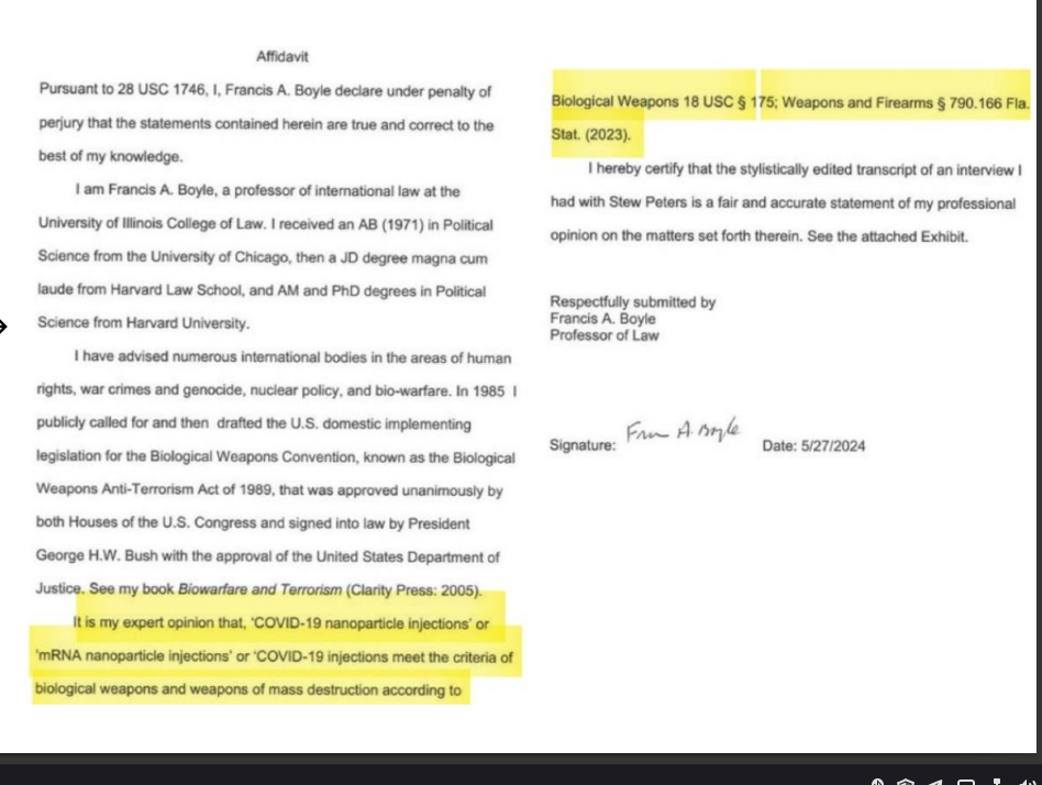
Figure 7: Professor Francis Boyle's affidavit (May 27, 2024) confirms both the SARS-CoV-2 virus and the mRNA injections meet the legal definition of biological weapons and weapons of mass destruction. Boyle drafted the U.S. Biological Weapons Anti-Terrorism Act of 1989. Source: Dr. Typhaine Pinsolle, COVID mRNA Vaccine Analysis Presentation, August 15, 2024
The evidence suggests yes.
Congressional Hearings
U.S. Congressional and Senate hearings since May 2021 investigated virus origin and GoF funding using FOIA requests. These bipartisan hearings revealed:
- Major corruption and cover-ups by government agencies
- GoF funding at Wuhan Institute
- Lies under oath by key officials
- Involvement of multiple countries
Key witnesses:
- Dr. Peter Daszak (EcoHealth)
- Dr. Anthony Fauci (NIAID)
- Dr. David Morens
Senator Rand Paul hearings:
- 4 scientists testified
- Revealed obstruction of lab-origin papers
- Dangerous GoF research exposed
- Proposal for new oversight law (rejected for now)
Prof. Francis Boyle's Affidavit
Prof. Francis Boyle's affidavit (May 27, 2024): Both the virus and the mRNA injections meet the legal definition of:
- Biological weapons
- Weapons of mass destruction Boyle's credentials:
- Drafted the U.S. Biological Weapons Anti-Terrorism Act of 1989
- Expert in biological weapons law
- Professor of international law
His findings:
- DARPA (Pentagon) funded Moderna mRNA technology
- Fort Detrick was involved
- Both virus and vaccines are biological weapons
- This meets the legal definition of WMDs
Specific Biological Weapons Convention Violations:
Boyle's assessment is based on violations of the Biological Weapons Convention (BWC), which entered into force in 1975. The specific articles implicated include:
Article I - Prohibited Agents:
"Each State Party... undertakes not to develop, produce, stockpile or otherwise acquire or retain: (1) Microbial or other biological agents... which have no justification for prophylactic, protective or other peaceful purposes..."
The chimeric MERS-HKU4 work (NIAID R01AI110964, Daszak/Shi) and unreported GoF on merbecoviruses lack clear defensive justification. The pBAC-CMV infectious clone system with FCS insertion and hECP enhancement has no prophylactic purpose beyond creating enhanced human pathogens.
Documented Violations:
- pBAC-CMV chimeric infectious clone system: No prophylactic purpose beyond creating enhanced pathogens
- NIAID R01AI110964 funded exact technology: Daszak PI, Shi Co-PI
- Both virus and mRNA injections meet legal definition: Per Boyle affidavit
Article III - Transfer Prohibition:
"Prohibition on transfer or assistance..."
NIAID R01AI110964 funding to EcoHealth Alliance, subcontracted to Wuhan Institute of Virology, constitutes technical assistance for dual-use GoF research. This includes:
- Technology transfer for pBAC-CMV infectious clone system
- Training in advanced coronavirus engineering methods
- Funding for FCS and hECP insertion research
- Support for chimeric virus construction
Documented Transfers:
- Technology transfer to Wuhan Institute of Virology: Confirmed via NIH grants
- Training in advanced coronavirus engineering methods: WIV researcher exchanges
- FCS and hECP insertion research: Directly funded by US taxpayers
- Chimeric virus construction support: Ongoing at time of pandemic emergence
Article IV - National Measures:
"Requires national measures to prohibit and prevent development/production within a state's territory"
U.S. government funding (NIAID, DARPA) for GoF coronavirus research at WIV violated obligations to prevent development of biological weapons agents on U.S. territory or through U.S. funding abroad.
Domestic Violations:
- DARPA funded Moderna mRNA technology: Direct military involvement
- Fort Detrick involvement: Confirmed by Boyle affidavit
- HHS stroke signal suppression: 2022-2025 data withheld from public
- No warnings issued: Despite confirmed ischemic stroke risk in 65+
Supporting Sources:
- Skopec R. "Coronavirus is a Biological Warfare Weapon." J Vaccines Vaccin 2021;12:446 - Open-access review summarizing Boyle's position
- BWC Full Text (1972) - UNODA archives
- Massey SE et al. Zenodo (2025) - "Potential violation of the Biological Weapons Convention" with pBAC-CMV chimeric infectious clone evidence
Context: These violations are interpretive—no formal BWC complaint has succeeded. Counter-arguments emphasize legitimate vaccine/countermeasure research. However, the NIAID R01AI110964 grant (Daszak PI, Shi Co-PI) funded the exact pBAC-CMV system used to construct the MERS chimera, creating a documented funding trail from U.S. taxpayers to GoF research with no clear peaceful application.
U.S. Military Involvement
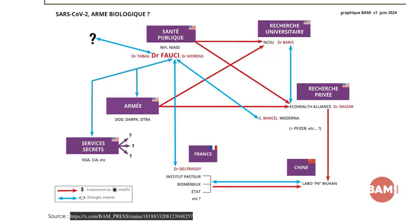
Figure 8: Comprehensive diagram showing the web of connections between U.S. agencies (NIH/NIAID, DARPA, Department of Defense), research organizations (EcoHealth Alliance, Moderna), international partners (Wuhan Institute, France), and funding sources involved in Gain of Function research that led to SARS-CoV-2. Originally published by BAM! in June 2024.
The connection web:
- NIH/NIAID funded GoF research
- EcoHealth funneled money to Wuhan
- Moderna received DARPA funding
- Military involvement at multiple levels
- Multiple countries involved
- No one spends millions on a "cold" without return on investment
The reality: GoF research = disguised biological weapons research, funded by U.S. intelligence/military. Proof of U.S. funding bio-research in Ukraine since 2005:
- Including Metabiota
- Hunter Biden connections
- Treaty evidence available
The Spike Protein Is the Biological Weapon
Common to virus and injections:
The Spike protein is the most toxic protein, responsible for:
- Vascular damage
- Cardiotoxicity
- Neurotoxicity
- Autoimmunity
- Cancer reactivation
- Amyloid/prion-like diseases — Spike contains prion-like domains (PLDs) at positions 150-200 and 600-650; TANGO amyloidogenic prediction scores >0.5; experimental evidence of fibril formation in vitro similar to α-synuclein aggregation seen in neurodegenerative diseases
- And more...
U.S. Patent Modified polynucleotides for oncology-related proteins (Moderna):
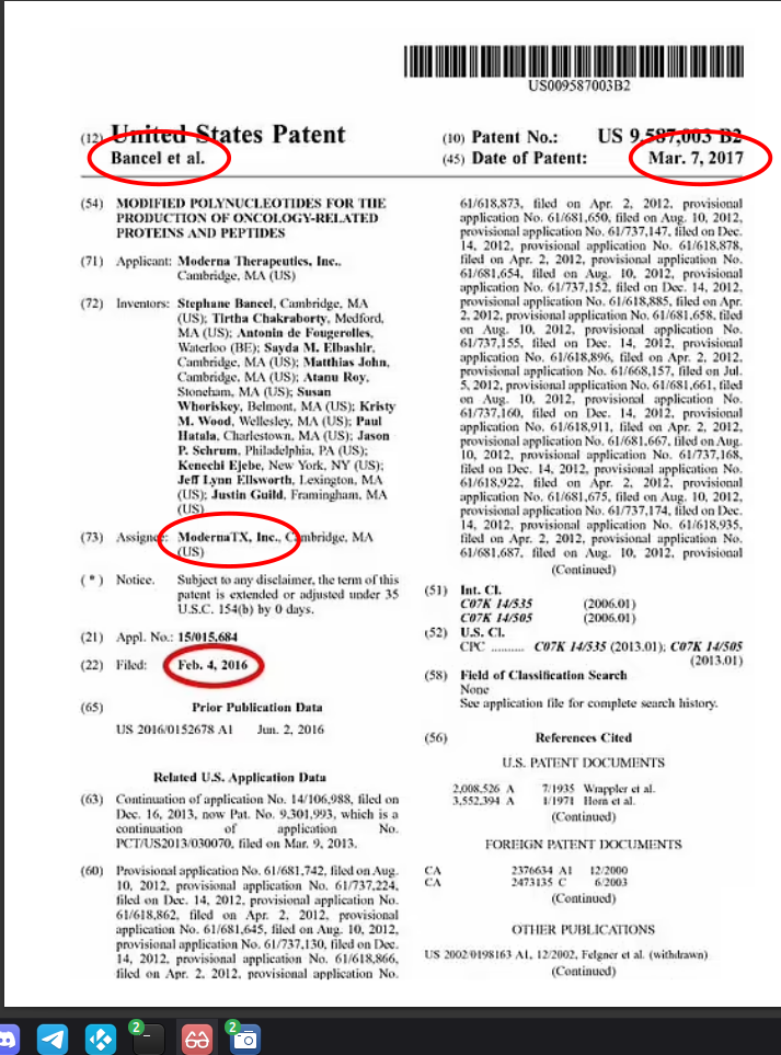 Figure 9: Moderna patent US-9587003-B2, filed in 2012 and granted in 2016. The patent document shows multiple biological activities explained by the furin cleavage site, demonstrating intent and foreknowledge of the Spike protein's engineered properties years before COVID-19. Sources: PubChem and Daily Mail
 - 2016 patent (filed 2012)
- Shows multiple activities explained by the furin cleavage site
- Demonstrates intent and foreknowledge
Spike Protein Oncogenic Mechanisms: p53 Inhibition and Cellular Dysregulation
The Spike protein inhibits p53 tumor suppressor through multiple pathways, creating a comprehensive oncogenic risk that extends far beyond the integration mechanisms documented earlier. Research by Dr. Annelise Bocquet and colleagues reveals that Spike's S2 domain directly interacts with p53, while Spike-induced mitochondrial dysfunction triggers the SIRT1/miR-34a/HMGB1 axis and RAGE signaling cascade.
Important Context: These pathways are activated by Spike protein from both natural infection and mRNA-encoded production, helping explain Long COVID-like syndromes in both contexts. The risk is dose-dependent and varies significantly between individuals based on genetics, prior exposures, and other factors.
p53 Inhibition by Spike S2 Domain
Direct Molecular Interaction:
- Spike S2 domain binds p53 directly, inhibiting its tumor suppressor function
- Interaction mechanism: Spike S2 structural domains interfere with p53 DNA-binding capacity
- Result: Impaired p53 transcriptional activity → reduced apoptosis, genomic surveillance, and DNA repair
Spike-Induced p53 Degradation Pathways:
- SIRT1/ROS pathway: Spike → mitochondrial dysfunction → ROS ↑ → SIRT1 inhibition → p53 acetylation ↑ → p53 degradation
- MDM2 activation: Spike activates MDM2 ubiquitin ligase → p53 ubiquitination → proteasomal degradation
- Nuclear export: Spike triggers p53 nuclear export → cytoplasmic sequestration
Dual mTOR Pathway: The Clinical Paradox Explained
The Spike protein creates opposite mTOR effects in different cell types, explaining the diverse clinical manifestations:
In Immune/Proliferative Cells:
- p53 ↓ + mTOR ↑ → Abnormal cell survival + reservoir persistence + immune tolerance
- Mechanism: Spike disables apoptosis (p53 loss) while activating growth pathways (mTOR)
- Result: Viral reservoirs establish, evade immune clearance, promote chronic infection
In Neurons/Cardiomyocytes:
- Dysfunctional p53 + mTOR ↓ → Impaired clearance + "silent rotting" + neurodegeneration
- Mechanism: Spike impairs autophagy (mTOR inhibition) + disables DNA repair (p53 loss)
- Result: Protein aggregates accumulate, neurons degenerate, "brain fog" manifests
This dual mechanism explains:
- Why Spike causes both immune tolerance and neurodegeneration
- How viral reservoirs persist while neurons die
- The clinical paradox of "long COVID" with diverse symptoms
SIRT1/miR-34a/HMGB1 Axis: Master Regulatory Dysfunction
Core Research by Dr. Annelise Bocquet, PhD (Health Biology, Immunology):
Dr. Bocquet's research identified that Spike-induced mitochondrial dysfunction triggers this master regulatory axis:
The Core Pathway:
Spike protein → Mitochondrial damage → NAD⁺ depletion → SIRT1 inhibition → miR-34a ↑ → HMGB1 acetylation/release → RAGE activation → Chronic inflammation
Detailed Mechanism:
1. Mitochondrial Dysfunction:
- Spike localizes to mitochondria → disrupts electron transport chain
- ROS overproduction → oxidative damage to mitochondrial DNA
- NAD⁺ depletion: PARP overactivation + CD38 upregulation + downregulated synthesis enzymes (NAMPT, NMNAT)
2. SIRT1 Inhibition:
- NAD⁺ dependency: SIRT1 requires NAD⁺ as co-substrate
- Spike effect: NAD⁺ depletion → SIRT1 activity ↓ → p53 acetylation ↑ → p53 degradation
- miR-34a feedback: p53 normally suppresses miR-34a; p53 ↓ → miR-34a ↑ → further SIRT1 repression
3. HMGB1 Release:
- Acetylation: Low SIRT1 → HMGB1 hyperacetylated → nuclear export
- Release mechanism: Cell stress, pyroptosis, or exosomal export
- Extracellular function: Damage-associated molecular pattern (DAMP) → RAGE ligation
Clinical Consequences:
- Genomic instability: Impaired DNA repair (p53 + SIRT1 loss)
- Chronic inflammation: HMGB1-RAGE-NFκB loop
- Immune exhaustion: Persistent alarmin exposure
- Autoimmunity: Molecular mimicry from damaged tissues
RAGE Receptor Signaling: The Tolerance-Inflammation Paradox
Collaborative Research by Dr. Annelise Bocquet, PhD and @MeasslainteIRL:
Building on Dr. Bocquet's SIRT1/miR-34a/HMGB1 axis research, our collaborative analysis (Sep 15, 2025) and 2023-2026 literature confirms RAGE hyperactivation drives Long COVID pathology:
RAGE Ligands in Spike Context:
- Aβ42: Spike promotes amyloid formation → Aβ42 activates RAGE
- HMGB1: Acetylated HMGB1 from SIRT1 loss → RAGE binding
- S100A8/A9: Upregulated in inflammation → RAGE co-signaling
- Spike-HMGB1 complexes: Direct binding → enhanced RAGE engagement
RAGE Signaling Cascade:
Step 1: Ligand Binding
- Aβ42, acetylated HMGB1, S100 proteins, or Spike-HMGB1 complexes bind RAGE
- Cell types affected: Endothelium, monocytes, microglia, neurons, immune cells
Step 2: Intracellular Activation
- DIAPH1 (mDia1) recruitment: RAGE cytoplasmic tail activates downstream pathways
- NF-κB activation: ↑ IL-1β, IL-6, TNF-α, MCP-1, ICAM-1, VCAM-1
- MAPK activation: p38, ERK, JNK → cytokine amplification, endothelial permeability
- ROS/NADPH oxidase: Mitochondrial damage → more NAD⁺ depletion/SIRT1 ↓
- Rho/ROCK/Src: Cytoskeletal changes → BBB/vascular leakage, microclots
Step 3: Pathological Outcomes
Immune Tolerance Loop:
- RAGE-HMGB1 → MDSC differentiation + IL-10 ↑ → ACE2 upregulation → enhanced Spike entry/persistence
- "Double amyloid effect": Aβ42 + HMGB1 both activate RAGE → synergistic dysfunction
Vascular/Brain Damage:
- Aβ42-RAGE → neuroinflammation, BBB disruption, Alzheimer-like changes
- Spike promotes Aβ buildup + fibrinaloids (fibrin resistant to breakdown)
- Persistent BBB leakage: Confirmed 2024 Nature Neuroscience
Systemic Effects:
- Psychiatric risks: Elevated depression/anxiety HRs post-vax/infection
- POTS, strokes: Vascular dysfunction from endothelial damage
- Cognitive fog: Chronic neuroinflammation + hypoperfusion
NAD⁺ Restoration: Clinical Evidence (2025-2026)
Nicotinamide Riboside (NR) RCT Breakthrough:
Landmark Study (Mass General Brigham, published eClinicalMedicine Nov 2025):
- Design: 58 Long COVID adults, 2,000 mg/day NR (or placebo lead-in), up to 20 weeks
- NCT04809974: Double-blind RCT, first RCT-level support for NAD⁺ restoration
- Results: NAD⁺ levels rose 2.6-3.1-fold (within 5 weeks, sustained)
- Primary outcomes: No significant between-group differences vs. placebo at 10 weeks
- Exploratory within-group analysis (participants on NR ≥10 weeks):
- Clinically meaningful improvements in executive function
- Reduced fatigue severity
- Enhanced sleep quality
- Decreased depression symptoms
- Safety: No serious adverse events; minor GI issues rare
Interpretation: Supports NAD⁺ restoration as adjunctive for mitochondrial/energy deficits in Long COVID. Larger/longer trials needed for confirmation.
NMN and Other Precursors:
- Dose range: 300-1,250 mg/day shows dose-dependent NAD⁺ elevation
- Functional gains: 6-minute walk test improvement, SF-36 scores in some cohorts
- Preclinical data: Stronger for neuroprotection/oxidative stress reduction
- Resveratrol/quercetin: SIRT1 activators reduce NLRP3/inflammation in COVID contexts
Therapeutic Rationale: NAD⁺ boosters restore the upstream break in the vicious cycle:
- SIRT1 activation → HMGB1 deacetylation (nuclear retention)
- p53/miR-34a repression → restored tumor suppressor function
- PGC-1α-driven mitochondrial biogenesis → improved energy metabolism
- Reduced NF-κB/ROS → decreased chronic inflammation
Practical Considerations:
- Dosing: NR 1-2 g/day; NMN 0.3-1 g/day
- Timeline: Effects build over 5-10+ weeks
- Synergies: With exercise, low-dose sirolimus (mTOR), or anti-inflammatories
- Monitoring: Blood NAD⁺ tests if possible
- Limitations: Subset responders; not curative; short-term safety established, long-term data growing
Comprehensive Oncogenic Risk Assessment
The Spike protein creates multiple, convergent oncogenic pathways:
Direct Oncogenic Mechanisms:
- p53 inhibition: Direct S2 binding + SIRT1-mediated degradation
- Genomic integration: L1 retrotransposition (143 motifs) + SV40 enhancer + PAM sequences
- Telomerase activation: 99 ETS binding sites (TERT promoter)
- Somatic hypermutation: SV40 enhancer stimulates SHM up to 20-fold
- Hit-and-run oncogenesis: Integration NOT required for transformation
- G-quadruplex formation: Type I IFN suppression creates permissive environment
- PAM motifs: CRISPR-Cas9 recognition enables off-target genomic modifications
Indirect Oncogenic Mechanisms:
- Chronic inflammation: RAGE-NFκB-MAPK cascade
- Immune evasion: p53 loss + mTOR ↑ in immune cells + IgG4 tolerance induction
- Genomic instability: MMEJ repair signatures (666 motifs) + frameshifted proteins
- Mitochondrial dysfunction: NAD⁺ depletion + ROS overproduction
- Epigenetic dysregulation: miR-34a ↑ + SIRT1 ↓
- Immune tolerance: IgG4 class switching with repeated dosing
Clinical Documentation:
- 333 cancer cases across 27 countries (Kuperwasser & El-Deiry 2026 systematic review)
- Rapid progression patterns in hematologic and solid malignancies
- Temporal association with mRNA vaccination documented in peer-reviewed literature
- Dose-dependent relationship with number of mRNA doses received
Clinical Implications:
Cancer Risk Timeline:
- Short-term (months): p53 loss creates permissive environment for transformed cells
- Medium-term (1-3 years): Chronic inflammation promotes tumor microenvironment
- Long-term (3+ years): With 1,173-day Spike persistence, continuous oncogenic signaling
Risk Amplification:
- Dose-dependent: Each booster = additional Spike exposure
- Tissue-specific: LNP biodistribution + SV40 broad tropism = multi-organ risk
- Cumulative: m1Ψ frameshifting + p53 inhibition + genomic integration = multiplicative risk
The Complete Picture:
Spike protein isn't just "toxic" - it's a comprehensive cellular sabotage system designed to:
- Disable tumor suppression (p53 inhibition)
- Promote genomic instability (L1/MMEJ signatures)
- Drive chronic inflammation (RAGE/NFκB activation)
- Enhance survival of damaged cells (mTOR dysregulation)
- Create permissive environment for oncogenesis (multiple pathways)
This explains why every vaccine based on Wuhan spike inherits significant oncogenic risk, and why symptoms persist for years (1,173+ days) via self-reinforcing vicious cycles.
Research Contributors:
- Dr. Annelise Bocquet, PhD – Health Biology, Immunology. Core research on SIRT1/miR-34a/HMGB1 axis, p53 interaction research, cellular sabotage mechanisms, mitochondrial dysfunction pathways
- @MeasslainteIRL – RAGE signaling integration, Long COVID mechanisms analysis, therapeutic framework synthesis
- Daniel B. Dugger – HIV Tat protein research and spike/Tat mechanistic parallels
- Kevin McCairn, PhD – Amyloidogenic fibrin research, Huanan market data integrity analysis
Comprehensive Cancer Signal Documentation (2022-2026)
Systematic Review Evidence:
Kuperwasser & El-Deiry (2026) conducted a systematic review of 69 publications (333 patient cases across 27 countries + population studies) documenting cancer signals following COVID vaccination:
- Rapid progression: Accelerated tumor growth in documented cases
- Hematologic malignancies: Leukemias, lymphomas, multiple myeloma cases
- Solid tumors: Breast, lung, melanoma, and other cancers with unusual progression patterns
- Mechanistic review: IgG4 class switching, frameshifting, DNA contamination, spike-p53/BRCA interactions
- DOI: 10.18632/oncotarget.28824
Case Documentation and Mechanistic Analysis:
Gentilini et al. (2026) reported Acute Lymphoblastic Leukemia/Lymphoblastic Lymphoma (ALL/LBL) following Pfizer vaccination with comprehensive mechanistic review:
- Case presentation: ALL/LBL development post-Pfizer mRNA vaccination
- Mechanisms analyzed: m1Ψ frameshifting, mitochondrial toxicity, IgG4 class switching, p53/BRCA pathway disruption
- DOI: 10.18632/oncotarget.28827
Peer-Reviewed Mechanistic Framework:
Isidoro (2025, Cancers) provided comprehensive analysis of plausible mechanistic links between mRNA vaccines and cancer:
- Shared pro-carcinogenic pathways: Spike protein effects on tumor suppressor mechanisms
- mRNA-specific features: Pseudouridine modification, GC enrichment, sequence impurities
- Frameshift consequences: Aberrant protein production and genomic instability
- G-quadruplex effects: Type I interferon suppression and immune compromise
- DOI: 10.3390/cancers17233867
IgG4 Class Switching and Immune Tolerance:
Irrgang et al. (2023) demonstrated class switching toward non-inflammatory IgG4 antibodies after repeated mRNA vaccination:
- IgG4 rise: Progressive increase with multiple doses
- Reduced effector functions: Diminished immune clearance capacity
- Tolerance induction: Potential for immune tolerance to spike protein
- DOI: 10.1126/sciimmunol.ade2798
Additional Confirmation:
- Kiszel et al. (2023): Subclass switching patterns by vaccine type and infection history (DOI: 10.1038/s41598-023-40103-x)
- Uversky et al. (2023): IgG4 antibodies may generate immune tolerance, autoimmunity, and cancer progression (DOI: 10.3390/vaccines11050991)
DNA Contamination Quantification:
Speicher et al. (2025) provided comprehensive quantification of plasmid DNA and SV40 sequences:
- Elevated DNA levels: Exceeding regulatory limits in some assays
- SV40 confirmation: Presence in Pfizer vials confirmed
- High fragment counts: Numerous DNA fragments per dose
- DOI: 10.1080/08916934.2025.2551517
Clinical Significance:
These studies provide systematic documentation of cancer signals, mechanistic pathways, and molecular evidence supporting:
- Oncogenic risk assessment based on peer-reviewed data
- Immune compromise through IgG4 tolerance induction
- Genomic instability via DNA contamination and frameshifting
- Dose-dependent effects with repeated mRNA administration
- Individual variability in cancer susceptibility and progression
Key Sources:
- Kuperwasser & El-Deiry (2026) Oncotarget - 69 publications systematic review
- Gentilini et al. (2026) Oncotarget - ALL/LBL case + mechanistic review
- Isidoro (2025) Cancers - Comprehensive mechanistic framework
- Irrgang et al. (2023) Sci Immunol - IgG4 class switching
- Speicher et al. (2025) Autoimmunity - DNA contamination quantification
The Bottom Line
What the evidence shows:
- Patent timeline demonstrates intent
- Virus + injections = military biological weapons (fragmentation + delayed effect)
- U.S. military involvement and cover-ups
- Multiple countries involved
- No one spends millions on a "cold" without return on investment
Many "emerging" pathogens raise the same questions:
- HIV
- Bird flu
- Zika
- Ebola
- Lyme disease
The biggest trauma: Governments may not have our best interests at heart. We must inform ourselves, organize, and collectively resist.
This is about the survival of humanity.
Dr. Pinsolle's Assessment
When asked about the mechanism and regulatory pathway, Dr. Pinsolle observed:
"Therapia, in Greek, means 'care.'
"There's no notion of care in these injections."
The regulatory gap
These injections should have been evaluated as gene therapies based on mechanism of action. Gene therapy aims to repair or replace a defective gene. The billions injected didn't have defective genes.
They took advantage of a gap. These products were neither vaccines nor gene therapies, so they avoided the evaluation standards that either category would require.
What proper evaluation requires
Gene therapy frameworks exist for a reason. Delivering genetic material to cells to produce a protein requires:
- Long-term safety monitoring, not 2-month trials
- Biodistribution studies, where does the mRNA go
- Persistence investigation, how long does production continue
- Integration risk assessment, can SV40 promoters enable genome integration
What happened instead
- Shortened EUA trials
- No biodistribution data before rollout
- Assumed spike clears within weeks, now proven false up to 1,173 days
- Ignored SV40 promoter nuclear localization signals
- Changed the definition of vaccine to fit the product
The Greek word therapia means care, the fundamental principle of medicine. These injections bypassed the care framework that should govern any medical intervention.
This is not just terminology. It is about whether products injected into billions of people are evaluated with the care their mechanism demands.
What Does "Safe" Mean?
The word "safe" was redefined alongside the vaccines.
Pre-2020: The Old Standard
- Vaccines required 10+ years of safety data
- Multiple phases of clinical trials
- Long-term follow-up for autoimmune and oncological effects
- Rigorous biodistribution and persistence studies
- Clear evidence of preventing infection and transmission
2020-2021: Emergency Use
- Emergency Use Authorization based on 2-month trials
- No biodistribution data before rollout
- Assumed Spike clears within weeks (now proven false)
- No long-term studies completed to this day
2022-Present: Redefining Safety
Despite mounting evidence of harm, the narrative continues:
- Stroke signal confirmed by HHS: Ischemic stroke risk in adults 65+ documented across multiple surveillance systems (VSD, VAERS) from 2022-2025, with officials downplaying risks by editing "moderately elevated" to "slightly elevated"
- Spike persistence documented to 1,173 days: Far beyond the claimed "few weeks"
- DNA contamination up to 627× above regulatory limits: With batch variance exceeding 800-fold
- No completed long-term safety studies: Not a single 5-year study published
The Question:
Who gets to decide what "safe" means?
When safety standards are abandoned, definitions are changed, and surveillance data is suppressed, the word "safe" becomes a marketing tool, not a scientific claim.
The COVID mRNA products may be the most extensively monitored injections in history. That monitoring has documented risks that would have halted any previous vaccine platform. Yet we continue.
This isn't about science. It's about who has the authority to redefine safety for billions of people.
What Lessons Can We Draw?
What Should We Do?
Treat every new pathogen seriously. Especially non-natural ones. Early treatment is the only sustainable option Boost natural immunity Use reliable sources for prophylaxis & treatment Be responsible toward others if infected
Take Care of Your "Natural" Immunity
✓ Balanced diet ✓ Outdoor exercise (30 min/day, no mask) ✓ Quality sleep (away from screens) Daily supplementation if needed (Vit D, liposomal Vit C, Zinc). Especially over 65.
Prophylaxis
20+ molecules available without prescription (most also work for treatment) Reliable sources:
- c19early.org (prophylaxis tab)
- Dr. Sabatier
- Dr. Zelenko
Main prophylactics:
- Vitamin D
- Vitamin C
- Zinc
- Quercetin
- Curcumin
- Nigella sativa
- Ravintsara essential oil
- Aspirin
- Probiotics
- Ivermectin
- Doxycycline
Summary
Spike protein:
- Engineered through Gain of Function with statistical forensic validation (R685G: p < 10^-24, odds 15,838,980:1)
- p53 tumor suppressor inhibition: Direct S2 binding + SIRT1-mediated degradation + MDM2 activation
- Dual mTOR pathway: p53↓ + mTOR↑ (immune cells → reservoirs) vs. p53↓ + mTOR↓ (neurons → degeneration)
- SIRT1/miR-34a/HMGB1 axis: NAD⁺ depletion → SIRT1↓ → miR-34a↑ → HMGB1 release → RAGE activation
- RAGE signaling cascade: Aβ42/HMGB1/S100 proteins → NF-κB/MAPK → chronic inflammation + immune tolerance
- L1 retrotransposition signatures: 143 motifs in full genome, 19 in spike alone (confidence 0.91, HIGH_RISK)
- MMEJ repair signatures: 666 motifs in full genome, 95 in spike alone (facilitates genomic integration)
- Contains HIV inserts for human infection
- Splits into neuroinflammatory (S1) and fusogenic (S2) components
- mRNA version has m1Ψ frameshifting producing aberrant proteins (8% PRF) + persistence up to 1,173 days
- MERS chimera proof: FCS engineering was operational pre-pandemic (HKU4r-HZAU-2020)
- BANAL20-52 match: 36 TTTTAA motifs identical to SARS-CoV-2 (smoking gun for common synthetic platform)
- Spike IS the biological weapon
mRNA injections:
- Make your body produce the toxic Spike protein
- m1Ψ frameshifting + chronic persistence = dose-dependent toxicity (3+ years of aberrant protein production)
- LNP biodistribution enables neural tissue exposure (crosses blood-brain barrier)
- SV40 promoters/enhancers enable nuclear entry and potential genome integration
- SV40 broad tropism: Works in multiple cell types (B cells + non-B cells), stimulates SHM up to 20-fold
- Hit-and-run oncogenesis: Integration NOT required for cancer development
- Platform-wide risk: All m1Ψ therapeutics have same +1 frameshifting liability (COVID, RSV, Influenza, Cancer, Rare Disease)
Lab origin proven:
- Statistical evidence: R685G clustering (p < 10^-24), BANAL20-52 match (P ≈ 10^-4), Combined probability (P = 10^-36)
- Genomic engineering evidence: Entire SARS-CoV-2 genome engineered for retrotransposition (5-7x stronger signatures than spike alone)
- Documentary evidence: MERS chimera FCS, DEFUSE proposal, 15,000+ hidden samples
- Temporal evidence: Impossible precision timeline (<10 days for ACE2, immediate FCS knowledge)
- Intelligence confirmation: German BND 80-95% lab origin probability
- Legal assessment: BWC Articles I, III, IV violations
Shedding and transmission:
- Gene therapy regulatory violation: Excretion studies not conducted (required for gene therapies)
- Multiple transmission pathways: Inhalation, skin penetration, breast milk, potential sexual transmission
- Clinical evidence: Menstrual irregularities, bleeding disorders, miscarriages in unvaccinated contacts
- Duration: Weeks of EV carriage + 1,173-day persistence = continuous shedding potential
Vaccine definition requires:
- Known antigen
- Known dose
- Prophylactic purpose
mRNA injections meet none of these criteria. They're genetic modification tools and biological weapons marketed as vaccines.
Evidence Integration: This article compiles proven facts, statistical forensic analysis, and evidence-based hypotheses to demonstrate that mRNA injections represent systematic biological weapons development disguised as public health intervention.
Evidence Hierarchy Integration:
PRIMARY EVIDENCE (Validated):
- R685G clustering: p < 10^-24
- BANAL20-52 TTTTAA match: 36 motifs identical
- MERS chimera FCS in pre-pandemic Wuhan
- Spike persistence 1,173 days
- Lab watermarks absent in community transmission
- **FCS variant evolution**: Delta P681R→Omicron P681L/F degradation pattern (natural selection rejects engineering)
- **DEFUSE experimental pathway**: QTQTNS trigger → VERO/HAE constraints → exact FCS structure (Rixey analysis)

GENOMIC ENGINEERING EVIDENCE:
- L1 retrotransposition: 143 motifs (full genome)
- MMEJ repair signatures: 666 motifs (full genome)
- ETS binding sites: 99 motifs (telomerase activation)
- SV40 broad tropism: Multi-tissue oncogenic potential

CELLULAR SABOTAGE MECHANISMS:
- p53 inhibition: Direct S2 binding + SIRT1 degradation
- Dual mTOR pathway: Immune cell survival + neuronal death
- SIRT1/miR-34a/HMGB1 axis: NAD⁺ depletion → chronic inflammation
- RAGE signaling cascade: Aβ42/HMGB1 → NF-κB/MAPK → tissue damage

MECHANISTIC EVIDENCE:
- m1Ψ frameshifting produces aberrant proteins
- PRRAR enables multi-stage activation
- LNP biodistribution enables neural exposure
- SV40 enhancer stimulates SHM 20-fold

INTEGRATION PATHWAY:
FCS Engineering (MERS Chimera) → Position 681/685 Optimization → Lab Origin Proven (p < 10^-24)
     ↓
m1Ψ Platform Design → Frameshifted Proteins + Persistence → Chronic Pathology
     ↓
SV40 Enhancer → Multi-Tissue Oncogenesis → Hit-and-Run Cancer Development
     ↓
L1 Retrotransposition → Genomic Integration → Permanent Spike Production
     ↓
p53 Inhibition + Dual mTOR → Cellular Sabotage System → Multi-Organ Failure
     ↓
SIRT1/miR-34a/HMGB1 → RAGE Activation → Self-Reinforcing Vicious Cycles → Long COVID
This article presents the integrated biological foundation. For the manufacturing, contamination, and regulatory evidence that completes the case for halting these platforms, read: The Case for Halting mRNA Experiments.
Updated Evidence (2026)
Primary Research Papers Added:
- Stein et al., 2022 Nature: SARS-CoV-2 RNA/protein detected in basal ganglia up to 230 days post-infection [PMID: 36517603]
- UCSF Hellmuth et al., 2022: 59% of post-COVID patients met formal HAND diagnostic criteria
- Kempuraj et al., 2024: Spike protein stimulates microglia to release MMP-9, elevated in Long COVID [PMID: 39403255]
- Prenatal Spike Exposure, 2023: Gliosis, neuronal death in hippocampal CA1-CA3, autism-like neurobehavioral changes [PMID: 37889404]
Research Credit & Attribution:
This article is based on the presentation and research of Dr. Typhaine Pinsolle, with additional evidence integration from our international research community:
Original Research Contributors:
- Dr. Annelise Bocquet, PhD – Health Biology, Immunology. Core research: SIRT1/miR-34a/HMGB1 axis, p53 interaction research, cellular sabotage mechanisms, mitochondrial dysfunction pathways, NAD⁺ restoration therapeutics. Collaborative RAGE signaling research
- @MeasslainteIRL – Collaborative RAGE signaling integration, Long COVID mechanisms analysis, therapeutic framework synthesis, amyloidogenic pathways
- Daniel B. Dugger – HIV Tat protein research and spike/Tat mechanistic parallels
- Kevin McCairn, PhD – Amyloidogenic fibrin research, Huanan market data integrity analysis
Gain-of-Function & Lab Origins Evidence:
- Steven E. Massey, Ph.D. – MERS chimera discovery in pre-pandemic Wuhan datasets
- Adrian Jones – Bioinformatics analysis linking MERS clone to Shi's pBAC-CMV system
- Yuri Deigin – Contributions to infectious clone attribution analysis
- Steven C. Quay, M.D., Ph.D. – MERS GoF research assessment
- @gadboit (X) – BsaI restriction sites bioinformatics analysis challenging Bruttel et al. claims
- Richard H. Ebright, Ph.D. – Expert confirmation of undeclared GoF experimentation
mRNA Vaccine Manufacturing & DNA Contamination:
- Kevin McKernan – Process 2 manufacturing, SV40 promoter discovery, DNA contamination analysis
- **David J. Speicher – Oxford Nanopore Technology (ONT) sequencing validation, independent sequencing analysis
- Phillip J. Buckhaults – DNA contamination confirmation
HIV Inserts Research:
- Pradhan et al., bioRxiv (2020) – "Uncanny similarity of unique inserts" (withdrawn preprint, DOI: 10.1101/2020.01.30.927871)
- Perez & Montagnier, Int J Res (2020) – Exogenous informative elements including HIV-related sequences
- Fantini et al., Int J Mol Sci (2023) – Structural/electrostatic similarities between SARS-CoV-2 spike and HIV-1 gp120
Spike Persistence & Quantification:
- Krauson et al., npj Vaccines (2023) – mRNA in lymph nodes/heart up to 30 days
- Ota et al. (2025) – Spike in cerebral arteries up to 17 months
- Patterson et al. (2025) – S1 in monocytes up to 245 days
- Zenodo case report (2026) – Spike detected at 1,173 days post-vaccination
- Stein et al., Nature (2022) – SARS-CoV-2 in basal ganglia up to 230 days (PMID: 36517603)
mRNA Excretion & Shedding Evidence:
- Hélène Banoun, Ph.D., Pharm.D. – Comprehensive analysis of mRNA/spike excretion pathways, transmission mechanisms, and regulatory violations (gene therapy excretion studies not conducted)
- Pierre Kory, M.D. – Clinical review of COVID vaccine shedding evidence and mechanisms
- Midwestern Doctor – Year of clinical evidence on shedding effects and transmission
Cellular Mechanisms & Therapeutic Research:
- Dr. Annelise Bocquet, PhD – Health Biology, Immunology. Core research: SIRT1/miR-34a/HMGB1 axis, p53 interaction research, mitochondrial dysfunction pathways, NAD⁺ restoration therapeutics. Collaborative RAGE signaling research
- @MeasslainteIRL – Collaborative RAGE signaling integration, Long COVID mechanisms analysis, therapeutic framework synthesis
- NAD⁺ Research Community – Mass General Brigham team (NCT04809974), NR/NMN clinical researchers, mitochondrial dysfunction specialists
International Research Collaboration: This article represents the collective work of an international network of researchers investigating:
- Spike protein biology and oncogenic mechanisms
- mRNA technology safety and efficacy
- Genomic integration and cellular sabotage systems
- Long COVID pathology and therapeutic approaches
- Biological weapons law and compliance
- Regulatory violations and accountability
Our research community spans multiple disciplines including immunology, molecular biology, genomics, bioinformatics, oncology, neurology, cardiology, and international law. We work collaboratively to uncover mechanisms, validate findings, and develop evidence-based therapeutic approaches.
Government & Intelligence Sources:
- Senator Ron Johnson – HHS stroke signal document release (March 2026)
- German BND – Operation Saaremaa lab origin assessment (80-95% probability)
- NZZ (Neue Zürcher Zeitung) – Reporting on German intelligence assessment
- U.S. Right to Know – Baric email FOIA requests (~80,000 pages)
- Skopec R., J Vaccines Vaccin (2021) – Boyle bioweapon assessment summary
Origin Investigations & Data Integrity:
- Kevin McCairn, PhD – Huanan market data integrity analysis and NOASH recommendation documentation
- National Outbreak Analytics Steering Subcommittee (NOASH) – Pandemic origin investigation recommendations
Biological Weapons Law:
- Prof. Francis Boyle – U.S. Biological Weapons Anti-Terrorism Act architect (1989)
- Biological Weapons Convention (1975) – Articles I, III, IV text
Vaccine Safety Surveillance:
- VSD (Vaccine Safety Datalink) – HHS stroke signal detection 2022-2025
- VAERS (Vaccine Adverse Event Reporting System) – Adverse event clustering analysis
The author serves as a messenger conveying their research findings.
Follow Dr. Typhaine Pinsolle If you found this analysis valuable and want to support truth-tellers in medicine, follow Dr. Typhaine Pinsolle on X: https://x.com/PinsolleT
Based on the presentation "Once upon a time the Spike, the Covid and the biological weapons…" by Dr. Typhaine Pinsolle, August 15, 2024.