Table of Contents
Key Takeaways
- In mice, mRNA-LNP vaccine immunity did not require direct dendritic-cell expression. The paper itself stresses this does not negate the importance of dendritic cells.
- Adding miR-122 target sites to detarget hepatocytes increased antigen-specific CD8+ T-cell responses about threefold in the same study.
- m1Ψ-substituted mRNA produces +1 frameshifted protein, about 8% of in-frame reporter levels. No adverse outcomes were reported in the human donors tested.
- Two autopsies detected vaccine-pattern spike in heart and liver with immune infiltration. Two cases cannot support any claim about frequency.
- Tissue-detargeting technology has existed since 2006 and was not used in the deployed vaccines. Whether it would have reduced cardiac risk is untested.
Correction, 4 October 2026. This article was first published 4 June 2026 with errors, corrected here against the source papers. The Marks paper was dated 2025; it was published 29 April 2026. Its senior author was attributed to the Weissman lab; the work is from Brian Brown's group at Mount Sinai. "Mört" was a misspelling; the first author is Mörz. The frameshifting rate was given as 0.5 to 1 percent; the paper reports frameshifted product at about 8 percent of in-frame reporter protein. The two autopsy findings were labelled "confirmed autoimmune myocarditis" and "confirmed autoimmune hepatitis"; the paper reports borderline histio-lymphocytic myocarditis in one case and chronic hepatitis in the context of pre-existing primary biliary cirrhosis in the other. The miRT origin paper was cited to Nature Biotechnology; it is Nature Medicine 2006. Overstated framing throughout was rewritten to what the papers show.
What this article covers#
Three peer-reviewed papers, read for what they establish and what they leave open:
- Marks et al., Nature Biotechnology, published 29 April 2026. Dendritic-cell expression and hepatocyte detargeting in mRNA-LNP immunity. Mouse data.
- Mulroney et al., Nature, December 2023. m1Ψ-substituted mRNA and +1 ribosomal frameshifting. In vitro, mouse, and small human sample.
- Mörz et al., Cells, May 2026. Vaccine-pattern spike protein in heart and liver tissue. Two autopsy cases.
All three concern vaccination. No infection-derived evidence is used or merged into the argument. Each is handled under its own evidence class: animal mechanism, molecular mechanism with a human blood sample, and case-report pathology.
What is actually shown#
Marks et al. 2026: dendritic-cell expression is not required, in mice#
Paper: Marks A, Siu S, et al. (senior author Brown BD, Icahn School of Medicine at Mount Sinai). "mRNA vaccine immunity is enhanced by hepatocyte detargeting and not dependent on dendritic cell expression." Nature Biotechnology, published 29 April 2026. Article · DOI 10.1038/s41587-026-03099-z
Population and assay. In vivo mouse models (C57BL/6, BALB/c, Ai14 reporter mice), mRNA-LNP formulated with the SM-102 lipid, delivered intravenously at 20 µg or intramuscularly at 5 µg, using reporter constructs and SARS-CoV-2 spike. miRNA target sites were inserted in the mRNA 3' UTR to switch expression off in specific cell types: 142T (hematopoietic cells, miR-142-3p), 122T (hepatocytes, miR-122), 133T/206T (myocytes, miR-133/miR-206). Readouts were flow cytometry of antigen-specific T cells, tissue-section imaging of transfected cells, and T-cell infiltration counts.
Findings, as the paper reports them:
- With a GFP reporter given intravenously, silencing hematopoietic expression (142T) reduced GFP-specific CD8+ T cells from roughly 10 percent to 5 percent, and silencing hepatocytes (122T) increased them to roughly 28 percent (their Figs 3c and 4b). The authors call the direction of the 122T result unexpected.
- With spike intramuscularly, the standard vaccine route, 142T made no difference to the T-cell response (their Fig 3d,e). Dendritic and other hematopoietic expression was dispensable for the response in this setting.
- Muscle cells can carry the response: transfected C2C12 myocytes transferred antigen to macrophages in co-culture, with roughly 12 percent of macrophages antigen-positive at 24 hours (Extended Data Fig 5g,h), and MHC-I was upregulated in transfected muscle in vivo.
- Liver was transfected even after intramuscular injection. In an adoptive-transfer model using highly reactive T cells (Jedi system), liver infiltration reached roughly 1,100 CD8+ T cells per mm² with unmodified RNA, and 122T prevented it (their Fig 5). The authors' own wording: "These results suggest that injection of RNA-LNPs can lead to killing of hepatocytes by antigen-specific T cells and this can be prevented by inclusion of 122T."
- Silencing myocytes (133T/206T) reduced spike-specific T cells after boosting by roughly 30 percent (their Fig 6i,j), so muscle expression contributed to, but did not carry alone, the response.
What the authors conclude, in their words: "This challenges a key assumption of mRNA vaccine effectiveness and is relevant for the design of mRNA therapeutics in which immunity is unwanted", and "Our findings do not negate the importance of DCs in mRNA vaccine immunity, but they do indicate direct mRNA expression in DCs is not necessary."
Limits. All efficacy data are mouse data. The authors state the translation limit themselves: "it is difficult to know how closely the functional biodistribution of mRNA-LNPs in animal models matches that in humans, as there is little characterization of the mRNA-LNP cellular expression patterns in humans." The hepatotoxicity readouts come from an adoptive-transfer model with deliberately reactive T cells, not from a standard vaccine regimen. Competing interests, same paragraph as the finding: senior author B.D.B. holds a patent on the use of miRNA target sites in gene vectors and consults for Merck. The detargeting result comes from the technology's holder.
The paper's own counter-statement, which belongs here rather than in a footnote: "It is important to note that, in clinical studies of RNA vaccines, overt liver and muscle toxicity has not been reported and we stress that they have proven to be very safe." The mouse mechanism stands. The clinical toxicity extrapolation is not the paper's claim, and this article does not adopt it.
Mulroney et al. 2024: m1Ψ causes +1 frameshifting#
Paper: Mulroney TE, Pöyry T, Yam-Puc JC, et al. (co-senior authors Thaventhiran JED, Willis AE, MRC Toxicology Unit, Cambridge). "N1-methylpseudouridylation of mRNA causes +1 ribosomal frameshifting." Nature 625:189-194, published online 6 December 2023. Article · DOI 10.1038/s41586-023-06800-3 · PMID 38057663.
Population and assay. Cell-free translation systems, HeLa cells, BNT162b2-vaccinated mice, and human blood samples: 21 BNT162b2 recipients and 20 ChAdOx1 recipients, none with reported undue effects. Readouts were mass spectrometry for frameshifted protein products and immune assays for reactivity to frameshifted peptides.
Findings, as the paper reports them:
- Replacing uridine with m1Ψ increased +1 ribosomal frameshifting. For a reporter construct, frameshifted product accumulated to about 8 percent of the corresponding in-frame protein (their Fig 1c). The 0.5 to 1 percent figure carried in the first version of this article was wrong and is withdrawn.
- Frameshifting mapped to specific slippery sequences. Mutating them synonymously eliminated detectable frameshifting, so this is a sequence-design problem, not an unavoidable property of the chemistry.
- Frameshifted products were detected in mice vaccinated with BNT162b2. Human BNT162b2 recipients showed immune reactivity to frameshifted peptides that ChAdOx1 recipients did not, in the sample tested.
What the authors conclude, in their words: "although there are no adverse outcomes reported from mistranslation of mRNA-based SARS-CoV-2 vaccines in humans, these data highlight potential off-target effects for future mRNA-based therapeutics and demonstrate the requirement for sequence optimization."
Limits. The human sample is small and was assayed for immune reactivity, not harm. The 8 percent figure is from a reporter construct, not the vaccine. Detection of frameshifted product is not evidence of pathology. Competing interests: T.E.M. and A.E.W. are inventors on a pending patent application related to mRNA technology.
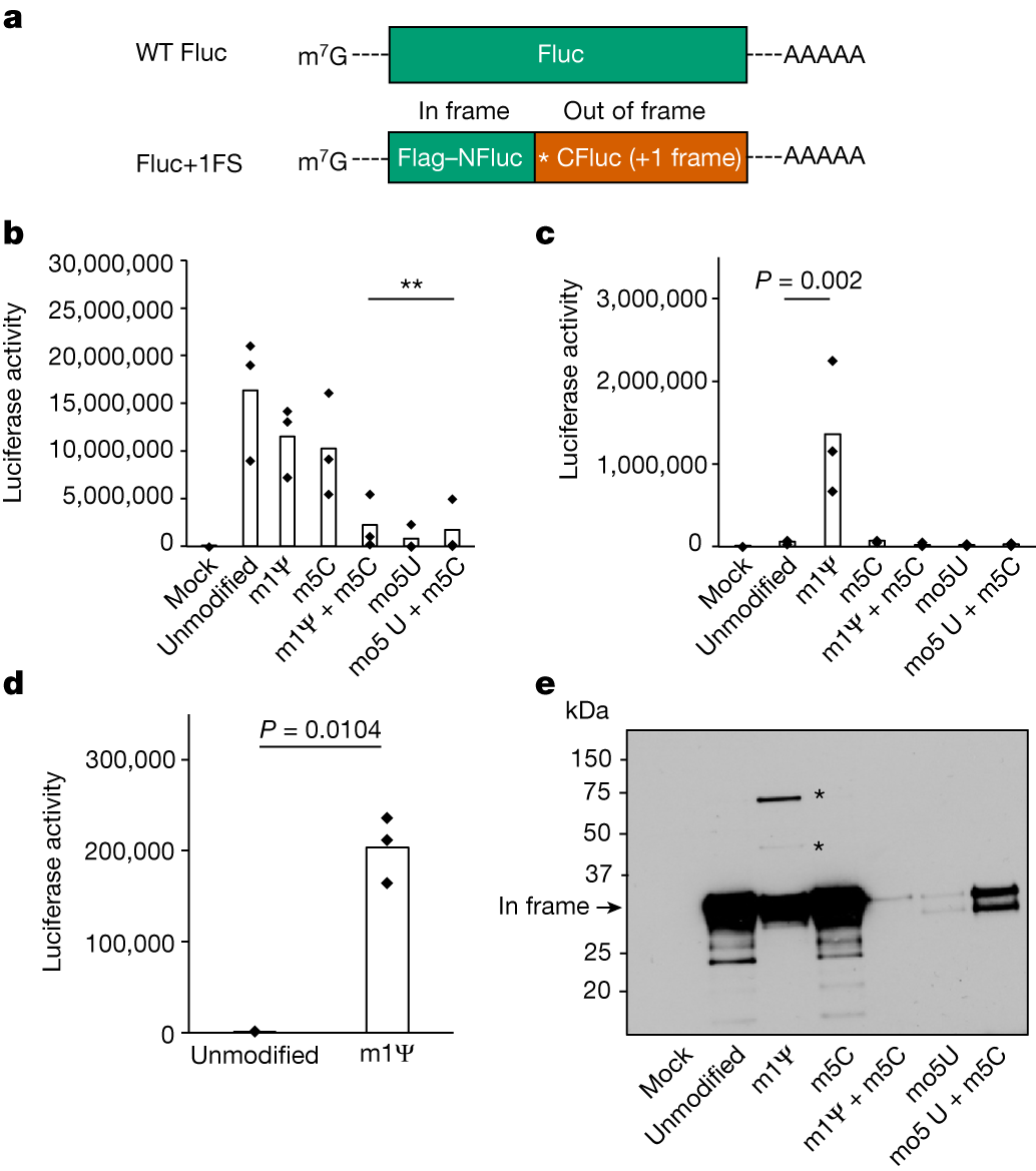 Figure 1: N1-methylpseudouridine causes +1 ribosomal frameshifting
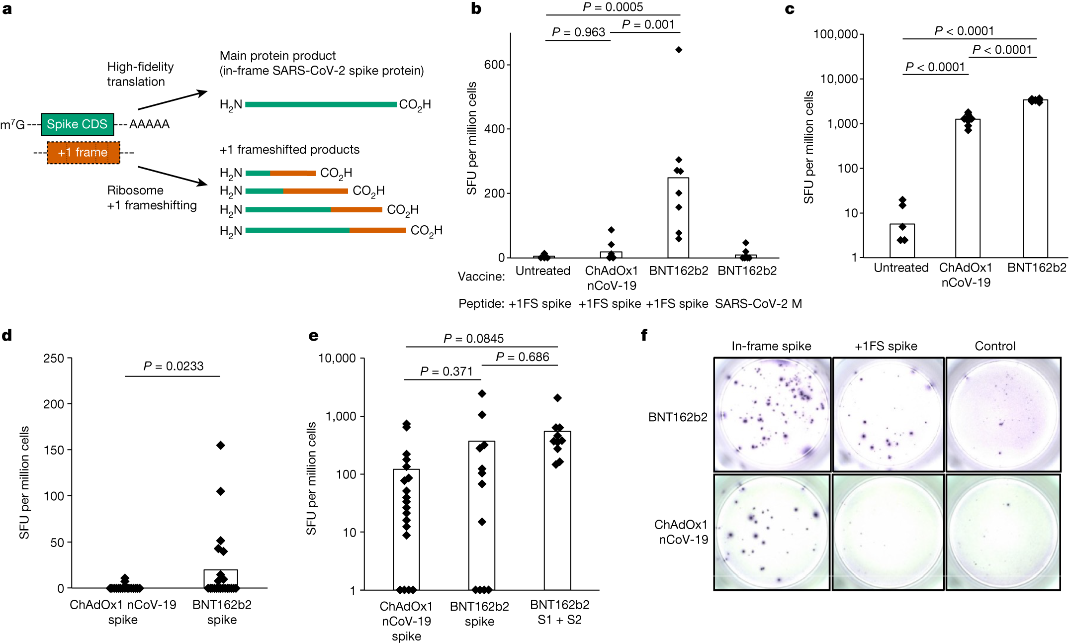 Figure 2: Frameshifted protein products
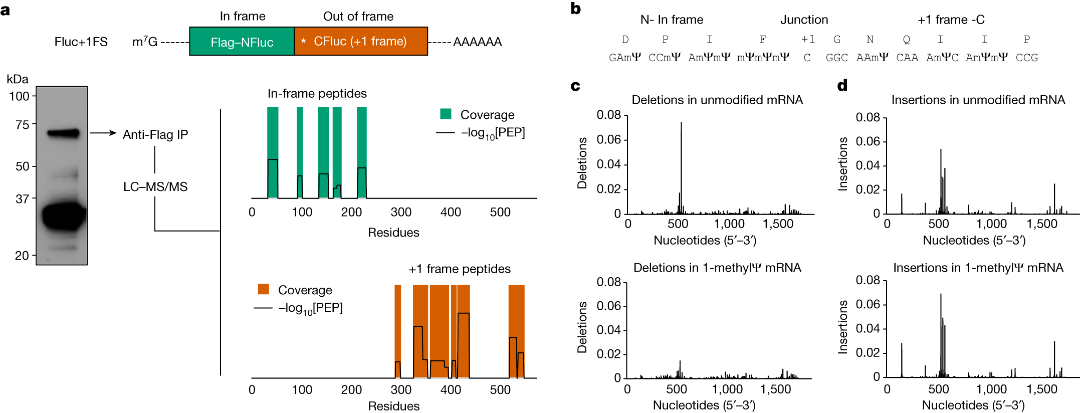 Figure 3: Mass spectrometry confirms frameshifted proteins
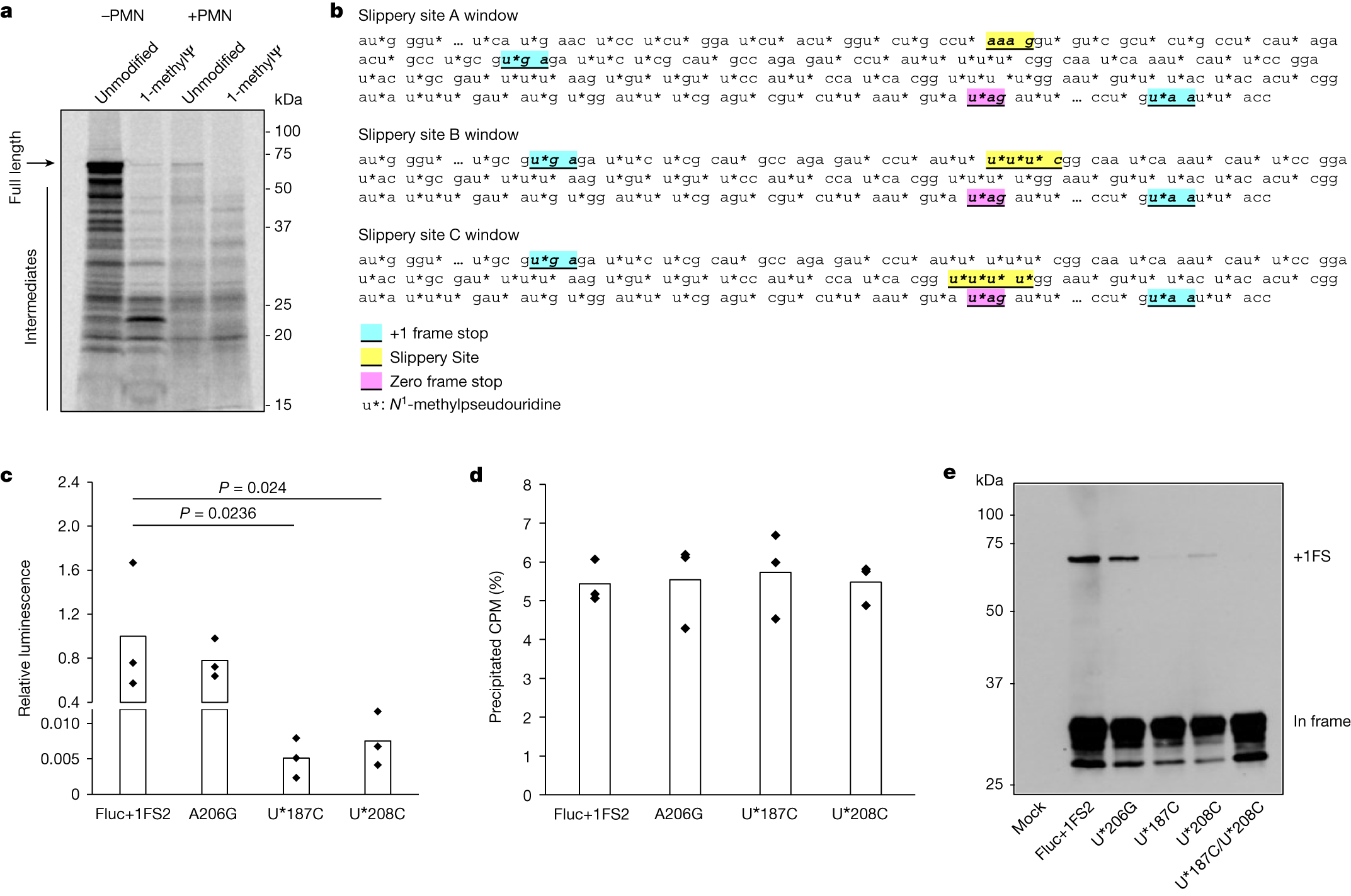 Figure 4: Quantification of frameshifted products
Mörz et al. 2026: two autopsy cases#
Paper: Mörz M, Donzelli A, Clancy RLC, Sano S, Fukushima M, Polykretis P. "Detection of Vaccine-Derived Spike Protein Associated with Immune Cell Infiltration in the Heart and Liver: A Report of Two Cases." Cells 15(11):978, published 26 May 2026. Article · DOI 10.3390/cells15110978
Population and assay. Two post-mortem cases, immunohistochemistry for spike subunit 1 and nucleocapsid, with CD4/CD8/CD68 immune-cell staining. Vaccine attribution rests on the spike-positive, nucleocapsid-negative pattern, compared against in-vitro vaccine-transfected control cells.
Case 1. A 72-year-old man who died of cryptogenic organizing pneumonia. History: two AstraZeneca doses, one Moderna, one Pfizer booster. Histology showed borderline histio-lymphocytic myocarditis. Spike subunit 1 stained positive in endothelial cells and infiltrating inflammatory cells. This is a report of detection with infiltration, not a finding of "confirmed autoimmune myocarditis"; the first version of this article said otherwise and is withdrawn.
Case 2. An 86-year-old who died of decompensated heart failure. History: three Pfizer doses. Histology showed chronic hepatitis in the context of primary biliary cirrhosis, a pre-existing autoimmune liver disease. Spike stained positive in CD68+ cells and sinusoidal endothelial cells. The authors state their own limit: "although double staining to confirm co-localization was not performed."
Limits. Two cases are case-report evidence. They show that the detection is possible. They cannot support any claim about how often it happens, and no such claim is made here. Competing interests, same paragraph as the finding: author P. Polykretis is an employee of Ancestralize Ltd.; the paper's discussion engages contested vaccine-policy questions. The staining methodology and the vaccine-attribution inference should be read with that in mind.
What is only suggested (labelled hypothesis)#
The design assumption#
The Marks paper's introduction states the prevailing assumption: mRNA-LNPs were understood to work primarily through uptake and expression in dendritic cells and other professional antigen-presenting cells. The new mouse data show direct dendritic-cell expression is not necessary there. That is a revision of a mechanistic account in one model system. It is not a demonstration that the platform "was built on a false foundation"; the clinical trials measured what the vaccines do, not the mechanism they were assumed to use. The mechanism was incompletely characterised, and the paper says so in the sentences quoted above.
Could cardiac detargeting have been used?#
The detargeting method is real and old. Brown et al. showed in 2006 that endogenous miR-142-3p target sites in a lentiviral vector suppressed transgene expression in hematopoietic lineages (Nature Medicine 12:585-591, PMID 16633348), and the group extended the approach to other tissues in 2007 (Nature Biotechnology 25:1457-1467, PMID 18026085). The deployed COVID-19 mRNA vaccines carry no miRNA detargeting sites. Marks et al. demonstrate the method working in the mRNA-LNP context for hepatocytes (122T) and myocytes (133T/206T) in mice.
This site's computational screen [COMP, hypothesis]. My own BLAST screen of the spike coding sequence found 31 perfect matches to cardiac-expressed miRNAs: 13 to miR-208a, 8 to miR-1, 5 to miR-133, 5 to miR-206, with example match positions at nucleotides 1143-1150 and 2183-2190. What this shows is sequence complementarity only. It does not show that endogenous cardiac miRNAs suppress spike translation in vivo; the screen did not test UTR context, miRNA abundance, or access. A detargeting construct would add designed target sites to the UTR, as in Marks, which is an engineering decision, not something the natural sequence does for you. Whether cardiac detargeting would have reduced myocarditis risk is untested in any system. The honest statement is: the method existed, the method works in mice, and it was not used. The counterfactual is unknown.
Does the Mörz pathology fit the Marks mechanism?#
Marks et al. show in mice that RNA-LNP expression in liver can draw antigen-specific T-cell killing, preventable by 122T. Mörz et al. report in two autopsies spike-positive cells with immune infiltration in heart and liver. The proposed sequence, that vaccine-derived expression in tissue attracts immune attack, is consistent across the two papers. Consistency is not confirmation. The papers use different species, different systems, and neither tests the other's claim. Proposed sequence; untested in humans.
Counter-evidence and what would falsify the claims#
Counter-Evidence & Limitations
Against the Marks extrapolation. The paper's own discussion states that overt liver and muscle toxicity has not been reported in clinical studies of RNA vaccines and that the vaccines "have proven to be very safe". Human cellular expression patterns for mRNA-LNPs are largely uncharacterised, so the mouse biodistribution may not match the human one. The senior author holds patents on the detargeting technology being validated.
Against frameshifting as a harm mechanism. Mulroney et al. found no adverse outcomes in their human donors, the 8 percent figure comes from a reporter construct rather than the vaccine sequence, and synonymous slippery-sequence redesign eliminates the effect. No study has connected frameshifted products to a clinical event.
Against the autopsy evidence carrying weight beyond two cases. Case reports cannot establish frequency, the co-localisation double staining was not performed, both patients had serious competing pathology, and vaccine attribution is an inference from a staining pattern rather than a sequencing result.
What would weaken or refute:
- Human mRNA-LNP expression mapping showing dendritic-cell expression is required after all, or that hepatocyte transfection after intramuscular injection is negligible in humans.
- Population-level pharmacovigilance and biopsy data showing no subclinical liver or muscle injury signal attributable to expression in those tissues.
- Failure to replicate the frameshifting results, or clinical studies showing frameshifted-product levels far below the reporter construct's.
- Broader autopsy series failing to reproduce the spike-positive, nucleocapsid-negative pattern, or reproducing it at rates matching infection-derived background.
Open questions the evidence leaves#
- What are the actual cellular expression patterns of mRNA-LNPs in humans, by route and formulation? The field's own answer is that this is largely uncharacterised.
- Would 122T or cardiac detargeting sites have changed the safety profile of the deployed vaccines? Untested in any species for cardiac endpoints; a mouse test is feasible and has not been run.
- What fraction of vaccine recipients made frameshifted products, at what levels, against what in-frame background?
- Does the Mörz staining pattern generalise? Answerable only by larger, blinded autopsy series with the double staining the authors themselves flag as missing.
- Regulators accepted a mechanism account that now needs revision in one model system. What expression-pattern data, if any, were filed? That is a documentary question, not a scientific one, and this article does not answer it.
Practical implications, stated at the strength of the evidence#
For future mRNA therapeutics, the papers' own implications are concrete: characterise expression patterns in humans rather than assuming them (Marks), and screen constructs for slippery sequences, since synonym choice removes the frameshifting (Mulroney). Detargeting sites are an available engineering option and were validated in the mRNA-LNP context in mice in 2026, twenty years after the underlying method was published.
For the deployed vaccines, no new clinical claim follows from these three papers. There is no randomised trial of detargeting, no established frameshifting harm, and no frequency estimate from two autopsies. The gap is real: a platform-level mechanism was revised in 2026 by a mouse experiment that could have been run years earlier. What that gap means for people who received the vaccines is exactly what is not yet established.
References#
- Marks A, Siu S, et al. "mRNA vaccine immunity is enhanced by hepatocyte detargeting and not dependent on dendritic cell expression." Nature Biotechnology, 29 April 2026. DOI 10.1038/s41587-026-03099-z
- Mulroney TE, Pöyry T, Yam-Puc JC, et al. "N1-methylpseudouridylation of mRNA causes +1 ribosomal frameshifting." Nature 625:189-194 (2024). DOI 10.1038/s41586-023-06800-3 · PMID 38057663
- Mörz M, Donzelli A, Clancy RLC, Sano S, Fukushima M, Polykretis P. "Detection of Vaccine-Derived Spike Protein Associated with Immune Cell Infiltration in the Heart and Liver: A Report of Two Cases." Cells 15(11):978 (2026). DOI 10.3390/cells15110978
- Brown BD, Venneri MA, Zingale A, Sergi Sergi L, Naldini L. "Endogenous microRNA regulation suppresses transgene expression in hematopoietic lineages and enables stable gene transfer." Nature Medicine 12(5):585-591 (2006). PMID 16633348 · DOI 10.1038/nm1398
- Brown BD, Gentner B, Cantore A, et al. "Endogenous microRNA can be broadly exploited to regulate transgene expression according to tissue, lineage and differentiation state." Nature Biotechnology 25(12):1457-1467 (2007). PMID 18026085 · DOI 10.1038/nbt1372