Table of Contents
Key Takeaways
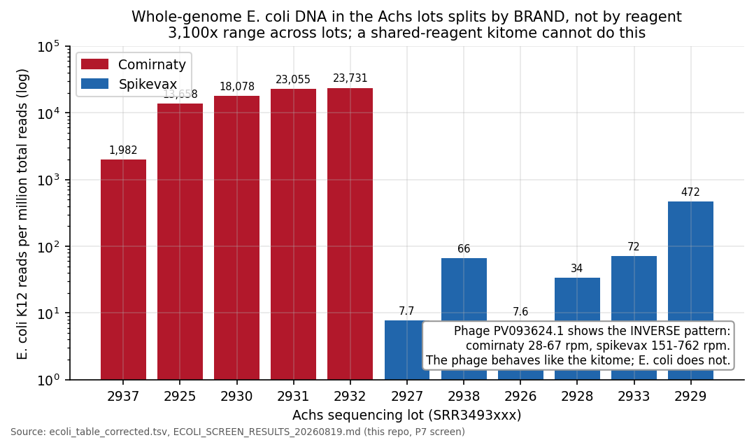
- The Achs paper's own sequencing deposit (PRJNA1303864) contains near-complete E. coli K12 genomic DNA in four of five Comirnaty lots: 1.4-2.4 percent of all reads (13,700-23,700 rpm) covering 96 percent of the genome at 56-95x mean depth. That refutes the paper's 'not from host genomes' conclusion.
- Whole circular plasmid is present in every lot; the circular fraction in Comirnaty is two to three orders of magnitude above the original-vector Spikevax lots.
- A fragment-length sign flip (+6 to +26 nt in backbone-continuous lots) matches the RNA:DNA hybrid protection mechanism.
Source documents. Achs et al., "Systematic analysis of COVID-19 mRNA vaccines using four orthogonal approaches demonstrates no excessive DNA impurities", npj Vaccines 10:259, 13 December 2025, DOI 10.1038/s41541-025-01304-9, with the sequencing deposit PRJNA1303864. The published method critique is McKernan, Speicher and Rose, "Systematic methodological flaws in DNA contamination assessment of mRNA vaccines: a critical analysis of Achs et al. (2025)", Oncotarget 2026;17:368-376. Every number in the reanalysis section below came off the public deposit with open tools, minimap2 and samtools.
TL;DR (1-minute read)
The method critique already stood on its own. The deposit adds this: the paper's own sequencing data contain near-complete E. coli genomic DNA in four of five Comirnaty lots, 1.4-2.4 percent of all reads (13,700-23,700 rpm) covering 96 percent of the genome at 56-95x mean depth, whole circular plasmid in every lot, and a fragment-length signature consistent with RNA:DNA hybrid protection, while the reported qPCR arithmetic converts a 12.2 ng over-limit figure into a reported 8 ng. The paper concludes the opposite of what its own data show, and it is cited to regulators as reassurance.
Every claim in the reanalysis section is checkable from PRJNA1303864 with public tools. The deposit is the authors' own.
Why this article exists#
On 13 December 2025, npj Vaccines published a paper whose title is its conclusion: no excessive DNA impurities. It has since been cited to regulators as reassurance that residual DNA in Comirnaty and Spikevax is within limits, the fragments are small, and nothing derives from host genomes.
The methodological critique of that paper already exists and is peer-reviewed, published in Oncotarget in August 2026 after the authors ignored the same points on the preprint server in September 2025 and a critique preprint in October 2025. That critique stands on its own. I do not repeat it here beyond a summary.
What I did was the boring, systematic version of the simplest test. Kevin's team had already used the deposit's coverage plots to expose the KAN-versus-spike qPCR problem, and as he put it, they got distracted by the rest of the technical pile before the Oncotarget paper went out. So I downloaded all fifteen runs Achs et al. deposited at NCBI under PRJNA1303864, re-mapped them, and checked every conclusion in the paper against the reads, lot by lot. This article is that pass.
What the paper claims, in its own words#
Two sentences carry the headline claims:
"Remaining reads in very low relative amounts either mapped to Escherichia coli K12 genome (GenBank accession NC_000913.3; non-pathogenic strain widely used in microbiology research and biotechnologies), bacteriophage discovered in microbiome study (PV093624.1...) or low complexity reads ... these reads can at least partially be a result of reagent contamination."
"83.52-98.08% of the DNA sequences were derived from plasmids used as templates during in vitro transcription of mRNA, rather than from exogenous sources or host genomes. These findings confirm that the detected DNA is a known and expected by-product of the manufacturing process, not an evidence of unintended contamination."
Read those two together. E. coli K12 is the production host of the DNA template plasmids. Reads mapping to it are host-genome reads by definition. The paper files them under reagent contamination and then concludes no host genomes. Both halves cannot be right, and the deposit decides which one is.
The method critique, summarised#
The full version is the Oncotarget paper and Kevin's article series. The eight points:
| # | The flaw | The effect |
|---|---|---|
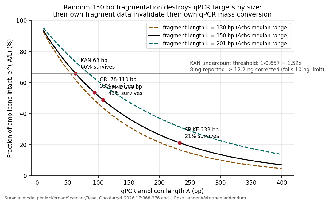
| 1 | qPCR amplicons sized 63 bp (KAN) to 233 bp (SPIKE) against a fragment population their own data put at a 130-201 bp median | The 233 bp spike target is mostly unamplifiable; the spike:backbone 4-6 Ct offset seen in four independent labs (McKernan, Speicher, Buckhaults, Fleming) disappears into a size artifact |
| 2 | Copy-to-mass conversion applied full-plasmid molecular weight to a fragmented single-target count | The only coherent readings are ~0.07 ng/dose (fragment mass) or 12.2 ng/dose (fragmentation-corrected, over the 10 ng limit). The published 8 ng sits between the two |
| 3 | No fragmentation correction anywhere in the qPCR chain | Even the 63 bp KAN amplicon needs a 1.5x correction at their own median fragment length; none was applied |
| 4 | Fluorometry switched to a DNA-purification front end no prior lab used, then the methods were edited post-hoc (ladder changed, no recovery number moved) | Recovery never validated by size, in a regime where kits lose sub-200 bp fragments preferentially |
| 5 | Capillary electrophoresis with electrokinetic injection, no matrix controls, standards in clean buffer | An LNP matrix that cannot inject reads as "below detection" regardless of true content |
| 6 | The CE instrument's smear floor is 50 pg/uL; the 10 ng per 300 uL dose limit equals 33.3 pg/uL | The instrument is blind across the entire band where compliance is decided |
| 7 | Fragment sizing done on Illumina after 95C x 10 min, two SPRI cleanups and 10 PCR cycles, in a lab that owns an ONT instrument | The measured "130-201 bp" medians describe the library prep, not the vials; ONT work from the same field shows kilobase fragments |
| 8 | Qubit trusted to titrate DNA input for their own Illumina runs, dismissed as unreliable when other labs reported over-limit DNA with it | Both statements appear in the same paper |
Jessica Rose's Lander-Waterman treatment gets to the same place as points 2 and 3 by a different road: detected KAN copies represent roughly 37 percent of the true KAN-bearing fragment population at conservative boundary assumptions, which pushes the corrected mass to around 25 ng per dose. Different boundary assumptions, same direction. Every coherent correction lands above the 10 ng limit.
Four things the discussion never touches: the 10 ng limit descends from naked-DNA kinetics and was never constructed for LNP-encapsulated DNA; Moderna's own patent US 10,077,439 B2 teaches against qPCR for this measurement; the same patent states residual DNA has the potential to be oncogenic; and Georgiou et al. 2006 documented PicoGreen underestimating fragmented DNA by up to 70 percent, which the fluorometry numbers inherit unexamined.
The 5lovak Academy of 5cience (McKernan's coinage)#
The section title is Kevin's joke, not mine, and it is doing serious work. The conflict-of-interest record below is his, assembled with receipts collected mostly by a researcher posting as Miroslav. I summarise it because the method critique and the deposit analysis cannot be read without it.
None of the COI material below is needed to refute the paper's conclusions. The methods and the deposit do that on their own.
The work was commissioned and paid for. Kevin documents approximately €350,000 paid for the study, funded through the Government of the Slovak Republic via the Ministry of Health. The commissioned institute is part of the Slovak Academy of Sciences, and its Biomedical Research Center entered a formal agreement with Sensible Biotech, a commercial mRNA company, on 28 August 2024, with shared laboratory infrastructure. Slovak government and EU investment in the institute's vaccine-capable infrastructure runs to roughly €25 million since 2014. The receipts are public: the institute's annual reports and the Slovak public-procurement register (crz.gov.sk) entries are linked from Kevin's article.
The record has since crossed into the press independently of Kevin. In August 2026, Epoch Times Slovakia re-reported it from the academy's own annual reports and the public-procurement register, naming the funder, the commercial partner and the institute's director (Milan Kajínek, 20 August 2026).
What that report adds, in English, for readers who do not read Slovak. Every item below is its reporting, anchored to the academy's own annual reports and the public-procurement register:
- The relationship is on paper in the academy's own annual reports. A cooperation agreement with Sensible Biotechnologies covering 2021 to 2023, valued at EUR 9,000, sits in the 2023 annual report. A further contract valued at EUR 4,500 sits in the 2024 report, signed by Silvia Pastoreková, director general of the Biomedical Research Center and a co-author of the study. The 2025 report lists the joint workplace with Sensible as opened.
- The joint workplace's contracted purpose, per the 2024 contract the report quotes, is analytical procedures for characterising mRNA samples. That is the same class of analytical work the study performs.
- The coordinator of the joint workplace is, per the report, Juraj Kopáček, director of the Institute of Virology and a main co-author of the study, who declared no conflict of interest for the paper. The joint-workplace contract itself is public in the procurement register (CRZ att/5241423): a 36-month cooperation agreement between the Biomedical Research Center and Sensible Biotechnologies, signed for the Center by Silvia Pastoreková, naming Juraj Kopáček as its contact person for contractual matters.
- The public-procurement register carries eleven contracts between Sensible Biotechnologies and the Ministry of Health and state institutions, including the Biomedical Research Center, across 2021 to 2026, which the report totals at more than EUR 28 million.
- Sensible Biotechnologies was founded in 2021 and has raised more than USD 13 million from investors including Y Combinator, BlueYard Capital and Onsight Ventures, the fund of a BioNTech co-founder.
- The Slovak Academy of Sciences did not respond to the outlet's request for comment by its deadline.
The problem is the dual role. The communicating author of the paper, who declared no conflicts of interest, is also, per Kevin's documentation, the institute director whose signature is on the collaboration agreement with the mRNA company. The conflict was raised in preprint commentary in September 2025, four months before publication. It is absent from the published paper.
What the COI record explains is why every methodological error in the paper points the same way: underestimation. A paper with eight independent biases that all err toward the sponsor's preferred answer is not a paper with eight unlucky accidents.
What the deposit itself shows#
I re-mapped eleven of the fifteen deposited libraries and extended the fragment-length comparison to all fifteen runs. What I ran: vector-unmapped reads re-mapped with minimap2 -ax sr against E. coli K12 NC_000913.3 (4,641,652 bp) and phage PV093624.1, primary alignments only, MAPQ >= 20; topology from full-depth proper-pair counts with a mid-reference control; depth with samtools depth -a -q 20 -d 0. Predictions went into the script headers before each run, including the ones that failed.
The fifteen deposited runs, with product, lineage and batch. "Re-mapped" is the eleven-library set above; "fragment lengths only" is the four runs added for the fifteen-run comparison:
| Run | Product | Lineage | Batch | Role here |
|---|---|---|---|---|
| SRR34932924 | Comirnaty | JN.1 | LK8456 | Fragment lengths only |
| SRR34932925 | Comirnaty | Original | not listed | Re-mapped |
| SRR34932926 | Spikevax | Bivalent BA.4/5 | 400012A | Re-mapped |
| SRR34932927 | Spikevax | Original | not listed | Re-mapped |
| SRR34932928 | Spikevax | Bivalent BA.1 | 200106A | Re-mapped |
| SRR34932929 | Spikevax | Bivalent BA.1 | 200023A | Re-mapped |
| SRR34932930 | Comirnaty | Original | 1L084A | Re-mapped |
| SRR34932931 | Comirnaty | Original | 1F1059A | Re-mapped |
| SRR34932932 | Comirnaty | Original | PCB0020 | Re-mapped |
| SRR34932933 | Spikevax | Bivalent BA.4/5 | MV1018A | Re-mapped |
| SRR34932934 | Comirnaty | KP.2 | LN0605 | Fragment lengths only |
| SRR34932935 | Comirnaty | JN.1 | LM9076 | Fragment lengths only |
| SRR34932936 | Comirnaty | JN.1 | LK8842 | Fragment lengths only |
| SRR34932937 | Comirnaty | Original | 1F1051A | Re-mapped |
| SRR34932938 | Spikevax | Original | 000058A | Re-mapped |
1. The host genome they said was not there#
In four of five Comirnaty lots (925, 930, 931, 932), 1.4-2.4 percent of ALL sequencing reads map across 96 percent of the complete E. coli K12 genome at 56-95x mean depth. That is 13,700 to 23,700 reads per million (rpm). All six Spikevax lots sit at 8-472 rpm. Every Comirnaty lot is above every Spikevax lot, the fifth Comirnaty lot (937) included: it sits seven to twelve times below its four siblings but still above every Spikevax lot. The 56-95x whole-genome depth figure describes the four high lots; the brand split holds across all five. The spread across brands is 3,100x.
Both brands were processed in the same lab with the same reagents and the same pipeline, and the vector-mapped loads overlap between brands (Comirnaty 106-127M vs Spikevax 80-115M reads). A shared-reagent kitome cannot produce a brand split. The phage PV093624.1 runs the other way (28-67 rpm in Comirnaty, 151-762 rpm in Spikevax): that is what an actual reagent component looks like, and it is uncorrelated with the E. coli signal.
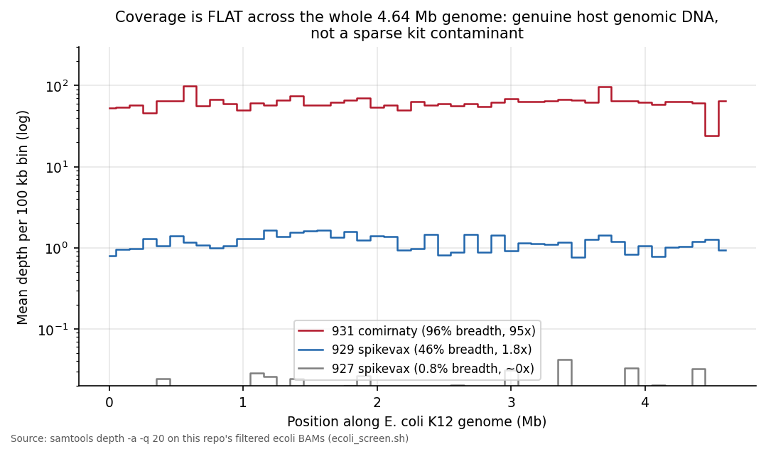
Coverage is flat along the entire 4.64 Mb genome in 100 kb bins. That is a genomic DNA population, not a sparse kit contaminant. This is manufacturing carryover, co-purified host genomic DNA from plasmid propagation, consistent in direction with Kevin's ONT finding of E. coli gDNA in Pfizer bivalent vials. "Rather than from ... host genomes" is refuted by the paper's own reads.
One more thing the paper's qPCR design guarantees: none of the assays target the E. coli genome at all. KAN, ORI and SPIKE are plasmid loci. A method that amplifies three plasmid windows is structurally blind to the entire host genome, which is the second reason, after the fragmentation bias in the method table, that only a universal double-stranded DNA measurement, Qubit after thorough RNase treatment in Kevin's framing, can see the total DNA in a vial.
2. Whole circular plasmid, not only small fragments#
Cross-junction read pairs, one read in the first 800 bp of the reference and the mate in the last 800 bp, proper pairs, MAPQ >= 20, full depth, mid-reference control, are nonzero in every lot. The circular molecule fraction, cross-junction pairs divided by mid-reference proper pairs from the same library, is ~1e-3 in all Comirnaty lots (1.4-2.7e-3: 3,472-6,964 cross-junction pairs against 2.1-2.6M mid-reference pairs) and the pv-vector Spikevax lots (1.0-9.7e-3), versus 7e-6 to 1.5e-5 in the two original-vector Spikevax lots (34 and 61 cross-junction pairs against 4.8M and 4.2M mid-reference). The pv-vector lots are the four bivalent Spikevax runs (926, 928, 929, 933), so named for the PV602126 bivalent vector they map to; the original-vector lots are 927 and 938.
Comirnaty coverage is flat across the whole plasmid and the T7 promoter is depressed, not peaked (T7:spike depth 0.58-0.77 in all five lots). The Spikevax enriched lots show the promoter at or above spike (1.03-1.54). Comirnaty spike:kanR sits at 0.43-0.64 across all nine Comirnaty runs in the corrected fifteen-run table, which reproduces the phase-2 numbers cross-platform.
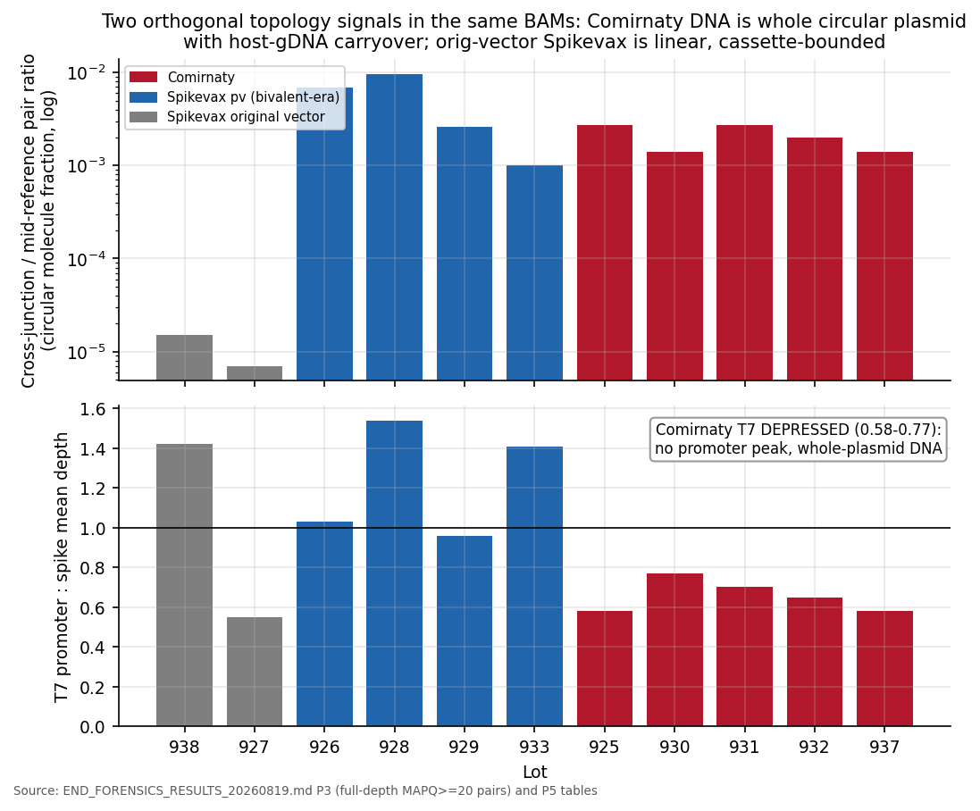
So the Comirnaty residual DNA is whole plasmid, with a circular fraction two to three orders of magnitude above the original-vector lots, plus host genomic DNA. It is the debris of an under-digested E. coli plasmid prep. The original-vector Spikevax lots are linear, cassette-bounded molecules. The paper's "small fragments" describe its own 95C x 10 min library prep, not the vials, and "small fragments only" is false for the paper's own Comirnaty data.
3. The RNA:DNA hybrid footprint, in their own fragment lengths#
Within-lot contrast, same library, same size selection: median spike-region fragment length minus median kanR-region fragment length. Every lot with continuous backbone coverage is positive, +6 to +26 nt. All nine Comirnaty runs sit at +6..+20 (sign test p = 0.004). The spike-rich pv lots 926 and 928 sit at +26 and +25. The three lots whose coverage collapses at the cassette boundary are the three negative ones (929/933 at -17, 938 at -21).
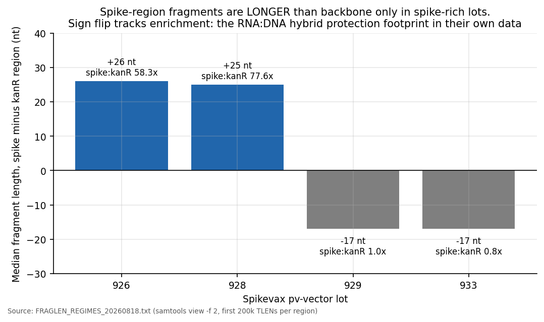
Longer, heavier spike-region DNA exactly where spike is enriched is what the hybrid-protection mechanism predicts: RNA:DNA-hybrid-protected spike DNA takes fewer DNase cuts. The mechanism and the prediction are McKernan, Rixey and Rose's, and BioNTech's own scientists published the hundredfold digestion-resistance of RNA:DNA hybrids (Lenk et al. 2024) a year before Achs. What I added is the measurement inside the Achs libraries. The 4-6 Ct spike enrichment that four independent labs measured, and that the Achs amplicon sizing erases, is sitting in the deposit's own fragment lengths.
Jessica Rose, co-author of both the Oncotarget critique and the RNA:DNA hybrids paper, put the finding this way:
During manufacturing of the nucleoside modified mRNA for the COVID-19 shots, the DNA template used was supposed to be destroyed by an enzyme, but the enzyme that the manufacturers selected (called DNaseI) does not chew up RNA:DNA hybrids - which inevitably would form as part of the process.
So RNA formed hybrids with the DNA that codes for spike during manufacturing. These hybrids protected the DNA from being cut so these protected (spike) pieces ended up longer on average than the unprotected backbone pieces - but only in the lots that still contained a lot of that spike DNA.
That is exactly the pattern shown in the graphs [above] - using the original authors' own sequencing data!
This is independent physical evidence that residual DNA in some of these vials was partially shielded by RNA:DNA hybrids.
BTW: The manufacturers have to have known both 1. that RNA:DNA hybrids would form (why wouldn't they, duh?) and 2. that DNaseI was the wrong enzyme to choose (also duh). Sutton et al. 1997, have published on this and so have Pfizer (Lenk et al., 2024)
THM: The Achs paper that we debunk in the attached paper (published in Oncotarget) shows precisely what we published in our RNA:DNA hybrids paper (also attached). If aliens came down and wanted to see a textbook case of situational irony and stupidity, I'd show them this.
The "attached paper" in her words is the Oncotarget critique and the RNA:DNA hybrids preprint, both linked in Sources. The plain-English mechanism she describes is the one the numbers in this section measure.
3a. The paper's own explanation, and why it does not hold#
The Achs authors saw the asymmetry themselves. Their Fig. 12 plots depth of coverage across the assembled plasmid genomes, and the text says: "Furthermore, genome coverage was significantly non-uniform across all samples, likely due to uneven residual DNA degradation (Fig. 12)." The sentence carries no citation, and the paper elsewhere leans on vial age to frame degradation, noting that "most of the vaccine batches were expired for an extended period". Two problems stand. Storage degradation has no mechanism that spares spike while destroying backbone: heat, nucleases and time act on the whole molecule, and no storage condition selectively deletes KAN. And the sequencing prep the authors themselves used, "Samples were first heated at 95 °C for 10 min to release nucleic acids from LNPs", fragments whatever DNA survived storage, uniformly, immediately before sequencing. A treatment the authors applied with their own hands overwrites any storage history. The asymmetry in their Fig. 12 was created after the vial, not in it, and its direction is the same one everything above measures: protected spike, unprotected backbone.
Kevin has quantified what that asymmetry is worth to a qPCR assay. Scanning the Achs reads for 100 bp windows, lowest-coverage regions in KAN against highest-coverage regions in spike, primer placement alone can swing the reported quantity 168x to 465x. An independent re-analysis of the same reads with 50 bp and 300 bp virtual amplicons, credited to Claude Science, lands at 5x to 615x. The starting observation on those coverage plots was mine, Kevin reproduced it, and the third-party analyses reached the same conclusion. Three chains, one direction: the paper's three-amplicon qPCR cannot arbitrate its own sequencing when primer placement alone moves the answer by two to three orders of magnitude.
4. The mixture scan, and why the brand split is not an artifact#
A minor-allele scan (>= 3 percent minor allele, MAPQ >= 20, baseQ >= 25, mpileup depth cap set above true depth) returns zero MIX rows in all five Comirnaty lots and both original-vector lots, and 29-68 MIX rows in every pv-vector lot, at the same shared spike positions with lot-varying fractions (926 near 50/50, 928/929/933 at 10-18 percent). A real second DNA population per pv lot, coherent within each lot.
The zero in every non-pv lot matters beyond the mixture finding: it bounds cross-lot contamination in the sequencing pipeline below 3 percent. That closes the standing objection that the E. coli brand split is index hopping or library carryover. It cannot be, because there is no cross-lot signal where a mixed-up pipeline would show one.
Standing caveats#
Sources#
- Achs et al. 2025, npj Vaccines 10:259: DOI 10.1038/s41541-025-01304-9
- Preprint: Research Square rs-7340318
- Sequencing deposit: PRJNA1303864
- McKernan, Speicher, Rose 2026, Oncotarget 17:368-376: full text
- October 2025 critique preprint: Zenodo 17410043
- Critical review: Zenodo 17409084
- RNA:DNA hybrids: Zenodo 17832183
- m6A / DAM methylation: Zenodo 17272427
- Lenk et al. 2024, Front Mol Biosci 11:1426129: DOI 10.3389/fmolb.2024.1426129
Press and public discussion:
- Epoch Times Slovakia on the COI record: Vedci SAV nepriznali prepojenie s výrobcami mRNA vakcín (Milan Kajínek, 20 August 2026, in Slovak; headline: "SAV scientists did not disclose ties to mRNA vaccine manufacturers, opponents claim")
- Discussion thread on the paper and the critique: ScienceGuardians
Kevin's article series on the Achs paper:
- CE method failures: More Holes in the Achs Paper
- Fluorometry prep standards: DNA Purification vs Direct Measurement
- DNaseI-XT witness experiment: Grok Gone Wild
- Nature Simps for Pharma
- Critiques etched into Bitcoin: Critical Review of Achs et al. (2025)
- RNA:DNA hybrids survive digestion: RNA/DNA Hybrids Survive Digestion
- Layman's Description of RNA/DNA Hybrids
- The COI record: The 5lovakian Academy of 5cience
David's article on the Achs work:
Open to corrections#
Further reading, sorry I know its a lot.
McKernan, K. (2026). RNase A reporting and citation completeness in assessments of residual DNA in mRNA vaccines. Zenodo. DOI: 10.5281/zenodo.22801568. ORCID: 0000-0002-3908-1122. Download PDF
McKernan, K. (2025). Hyperstimulatory N⁶-methyladenine (m6A) in residual SV40 plasmid DNA in mRNA vaccines. Zenodo. DOI: 10.5281/zenodo.17281691. ORCID: 0000-0002-3908-1122. Download PDF
Every number here can be reproduced from PRJNA1303864 and every claim carries its source. If I have something wrong, tell me and I will correct it, and the correction gets the same prominence as the original claim. If the authors want to respond point by point, I will publish their response here, unedited. Standing offer, no expiry. General policy on the Methodology page.